. 1998 Nov 2;143(3):827-36.
doi: 10.1083/jcb.143.3.827.

TGF-beta plays a key role in morphogenesis of the pancreatic islets of Langerhans by controlling the activity of the matrix metalloproteinase MMP-2

Affiliations

TGF-beta plays a key role in morphogenesis of the pancreatic islets of Langerhans by controlling the activity of the matrix metalloproteinase MMP-2

F Miralles et al. J Cell Biol. .

Abstract

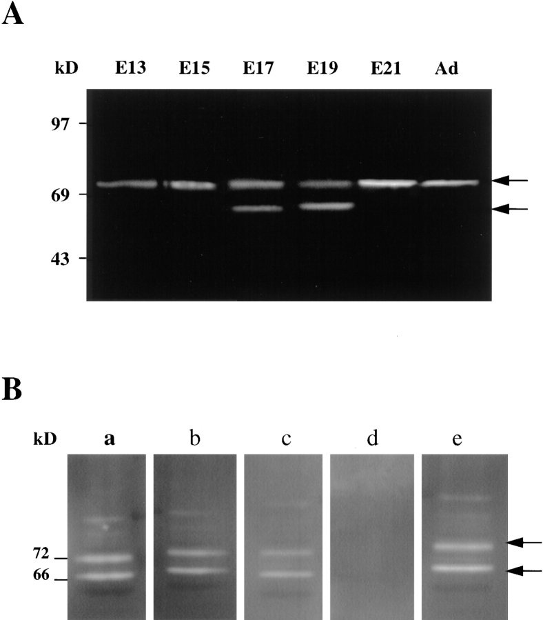
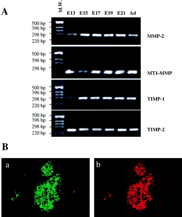
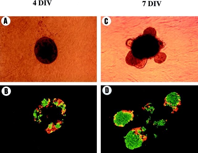
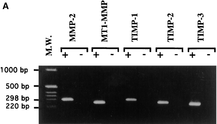
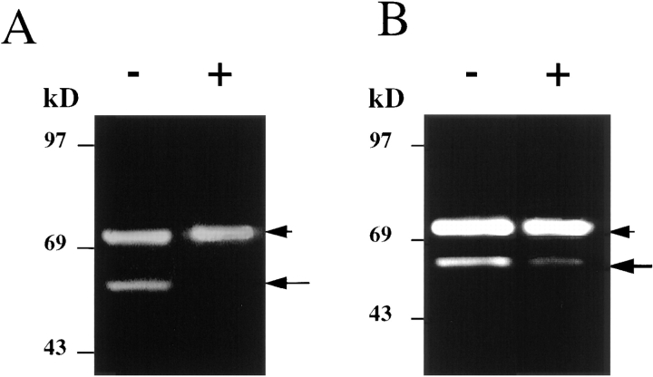
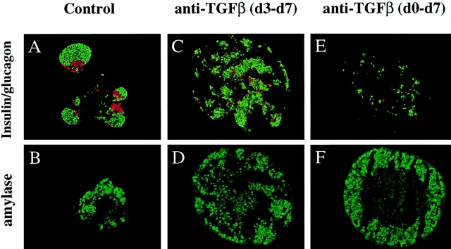
Islets of Langerhans are microorgans scattered throughout the pancreas, and are responsible for synthesizing and secreting pancreatic hormones. While progress has recently been made concerning cell differentiation of the islets of Langerhans, the mechanism controlling islet morphogenesis is not known. It is thought that these islets are formed by mature cell association, first differentiating in the primitive pancreatic epithelium, then migrating in the extracellular matrix, and finally associating into islets of Langerhans. This mechanism suggests that the extracellular matrix has to be degraded for proper islet morphogenesis. We demonstrated in the present study that during rat pancreatic development, matrix metalloproteinase 2 (MMP-2) is activated in vivo between E17 and E19 when islet morphogenesis occurs. We next demonstrated that when E12.5 pancreatic epithelia develop in vitro, MMP-2 is activated in an in vitro model that recapitulates endocrine pancreas development (Miralles, F., P. Czernichow, and R. Scharfmann. 1998. Development. 125: 1017-1024). On the other hand, islet morphogenesis was impaired when MMP-2 activity was inhibited. We next demonstrated that exogenous TGF-beta1 positively controls both islet morphogenesis and MMP-2 activity. Finally, we demonstrated that both islet morphogenesis and MMP-2 activation were abolished in the presence of a pan-specific TGF-beta neutralizing antibody. Taken together, these observations demonstrate that in vitro, TGF-beta is a key activator of pancreatic MMP-2, and that MMP-2 activity is necessary for islet morphogenesis.


Figures

Figure 1
Figure 1
Metalloproteinases activity in the developing pancreas: (A) Equal amounts of protein extracts from each of the stages indicated were analyzed by zymography. A proteinase of 70–72 kD, representing the proform of the enzyme MMP-2, was detected in samples of all the stages of pancreatic development. The 62–64 kD activated form of the enzyme is essentially detectable between E17 and E19. (B) Protein extracts from E19 rat pancreas were run on a 8% SDS-polyacrylamide gel copolymerized with 1 mg/ml of gelatin. The gel was next divided into sections for subsequent overnight digestion at 37°C in incubation buffer (50 mM Tris, 10 mM CaCl2, pH 7.6) containing (a) no additive; (b) 10 μM BB-3103; (c) 50 μM BB-3103; (d) 1 mM compound BB-3103; or (e) 0.1 μl/ml DMSO diluent.
Figure 2
Figure 2
Expression of matrix metalloproteinase components in the pancreas in development. (A) RT-PCR analysis of MMP-2, MT1-MMP, TIMP-1, and TIMP-2 expression. First-strand cDNA was synthesized and used as template in PCR reactions. 30 cycles of amplification were performed for each reaction. (B) Sections of newborn rat pancreas were costained with (a) anti-insulin and (b) anti-MMP-2 antibodies.
Figure 3
Figure 3
Morphological and immunohistological analysis of pancreatic rudiments developed in vitro. Pancreatic rudiments devoid of their surrounding mesenchyme were grown into tridimensional collagen gels. Pancreatic rudiments were photographed (A and C) and analyzed by immunohistochemistry (B and D) for insulin (green) or glucagon (red) expression after 4 (A and B) and 7 d (C and D) of culture.
Figure 4
Figure 4
Expression of the matrix metalloproteinases components in pancreatic rudiments in culture. (A) RT-PCR analysis of MMP-2, MT1-MMP, and TIMPs 1–3 expression in pancreatic rudiments cultured for 4 d. First-strand cDNA was synthesized and used as template in PCR reactions. 30 cycles of amplification where performed for each reaction. (B) Immunohistochemical analysis of insulin and MMP-2 expression in pancreatic rudiments. Sections were costained with (a) anti-insulin and (b) anti-MMP-2 antibodies.
Figure 4
Figure 4
Expression of the matrix metalloproteinases components in pancreatic rudiments in culture. (A) RT-PCR analysis of MMP-2, MT1-MMP, and TIMPs 1–3 expression in pancreatic rudiments cultured for 4 d. First-strand cDNA was synthesized and used as template in PCR reactions. 30 cycles of amplification where performed for each reaction. (B) Immunohistochemical analysis of insulin and MMP-2 expression in pancreatic rudiments. Sections were costained with (a) anti-insulin and (b) anti-MMP-2 antibodies.
Figure 5
Figure 5
Compound BB-3130 inhibits islet morphogenesis without affecting cell differentiation. The rudiments were grown for 7 d in the absence (control) or in the presence of BB-3103 (1 μM or 10 μM). Adding 1 μM or 10 μM of compound BB-3103 inhibits islet morphogenesis (compare A to B and C). On the other hand, immunohistochemical analysis revealed that differentiation into insulin-positive cells in green or into glucagon-positive cells in red occured both in the absence (D) or in the presence (E and F) of inhibitor. The differentiation into amylase-positive cells did also occur in the absence (G) or in the presence (H and I) of BB-3103.
Figure 6
Figure 6
Morphological and immunohistological analysis of pancreatic rudiments developed in vitro in the presence of exogenous TGF-β1. Pancreatic rudiments devoid of their surrounding mesenchyme were grown into tridimensional collagen gels in the absence (A, C, E, and G) or in the presence (B, D, F, and H) of TGF-β1. Pancreatic rudiments were photographed and analyzed by immunohistochemistry for insulin (green) or glucagon (red) expression after 4 (A–D) and 7 d (E–H) of culture. Note that after 4 d in culture in the presence of TGF-β1, translucent buds were already emerging (B), and many endocrine cells showed a spindled shape characteristic of cells that are in migration (D), which was not the case after 4 d in the absence of exogenous TGF-β1.
Figure 7
Figure 7
Analysis of gelatinase expression and activity in pancreatic rudiments cultured in the presence of exogenous TGF-β1. Pancreatic rudiments grown for 4 or 7 d in the presence or absence of TGF-β were analyzed for MMP-2 activity by gel zymography. Lanes 1 and 2: pancreatic rudiment grown for 4 d without (lane 1) or with (lane 2) 1 ng/ml TGF-β1. Lanes 3 and 4: pancreatic rudiment grown for 7 d without (lane 3) or with (lane 4) 1 ng/ml TGF-β1.
Figure 8
Figure 8
Expression of TGF-β and its receptors in pancreatic rudiments in culture. cDNAs were prepared from pancreatic rudiments devoid of mesenchyme and cultured for 4 d in collagen gels. Then they were used as template in PCR reactions with specific primers for TGF-β1, TGF-β2, TGF-β3, TGF-βRI, and TGF-βRII. (A) 30 cycles of amplification. (B) 35 cycles of amplification.
Figure 9
Figure 9
Analysis of gelatinase A expression and activity in pancreatic rudiments grown in the presence of a panTGF-β neutralizing antibody. The pancreatic rudiments were cultured in the presence of a panTGF-β neutralizing antibody, and after a 7-d culture period, the rudiments were recovered and their gelatinase activity was analyzed by gel zymography. The antibody was added either from the third (A) or from the first (B) day of culture. (−) rudiments cultured in the presence of nonimmune serum; (+) rudiments cultured in the presence of the TGF-β neutralizing antibody.
Figure 10
Figure 10
Analysis of pancreatic rudiments grown in the presence of a panTGF-β neutralizing antibody. To define whether TGF-β is necessary for islet formation, pancreatic rudiments were cultured in the presence of a pan-specific TGF-β neutralizing antibody. The antibody was added either from the first (d0) or the third (d3) day of the culture. The rudiments were analyzed by immunohistochemistry for insulin, glucagon, and amylase expression. In the control rudiments, the insulin- (green) and glucagon- (red) expressing cells are arranged into islet-like structure (A), budding from the core of the rudiment that is mainly composed of amylase-expressing cells (B). When the rudiments are cultured in the presence of the panTGF-β neutralizing antibody from the third day of culture (d3–d7), the endocrine cells differentiate, but do not bud (C), and form small clusters interspersed between the acinar cells (D). When the rudiments are cultured in the presence of the panTGF-β neutralizing antibody from the first day of culture (d0–d7), a more drastic effect is observed. The endocrine cells appear dispersed (E) and surrounded by acinar cells (F).

References

    1. Ahlgren U, Pfaff S, Jessel T, Edlund T, Edlund H. Independent requirement for ISL1 in formation of pancreatic mesenchyme and islet cells. Nature. 1997;385:257–260. - PubMed
    1. Ancuta P, Fahma H, Pons J, LeBlay K, Chaby R. Involvement of the membrane form of tumour necrosis factor-alpha in lipopolysaccharide-induced priming of mouse peritoneal macrophages for enhanced nitric oxide response to lipopolysaccaride. Immunology. 1997;92:259–266. - PMC - PubMed
    1. Atouf F, Czernichow P, Scharfmann R. Expression of neuronal traits in pancreatic beta cells: implication of NRSF/REST, a neuron-restrictive silencer. J Biol Chem. 1997;272:1929–1934. - PubMed
    1. Bottinger E, Jakubczak J, Roberts I, Mumy M, Hemmati P, Bagnall K, Merlino G, Wakefield L. Expression of a dominant-negative mutant TGF-β type II receptor in transgenic mice reveals essential roles for TGF-β in regulation of growth and differentiation in the exocrine pancreas. EMBO (Eur Mol Biol Organ) J. 1997;16:2621–2633. - PMC - PubMed
    1. Chin J, Werb Z. Matrix metalloproteinases regulate morphogenesis, migration and remodeling of epithelium, tongue skeletal muscle and cartilage in the mandibular arch. Development. 1997;124:1519–1530. - PubMed

Publication types