. 1998 Nov 2;143(3):861-73.
doi: 10.1083/jcb.143.3.861.

Impaired integrin-mediated adhesion and signaling in fibroblasts expressing a dominant-negative mutant PTP1B

Affiliations

Impaired integrin-mediated adhesion and signaling in fibroblasts expressing a dominant-negative mutant PTP1B

C O Arregui et al. J Cell Biol. .

Erratum in

  • J Cell Biol 1998 Dec 14;143(6):1761

Abstract

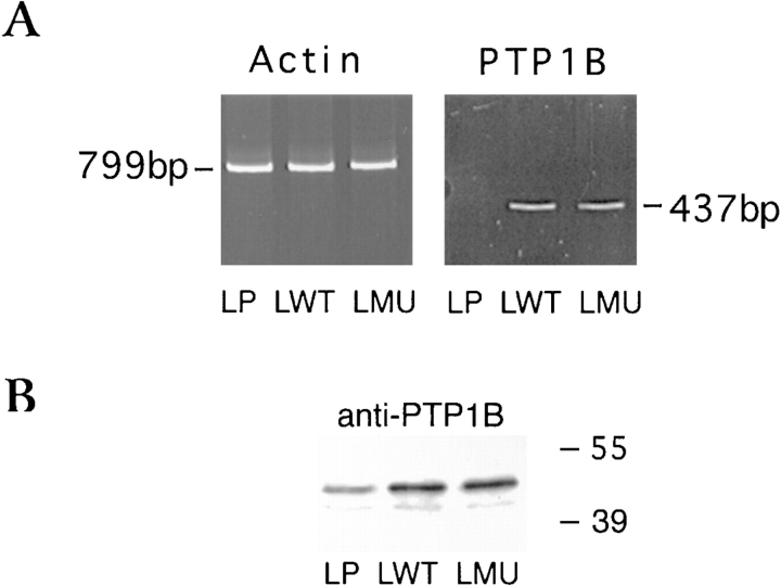
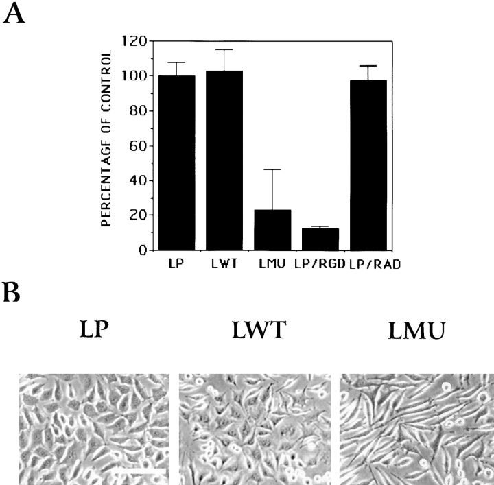
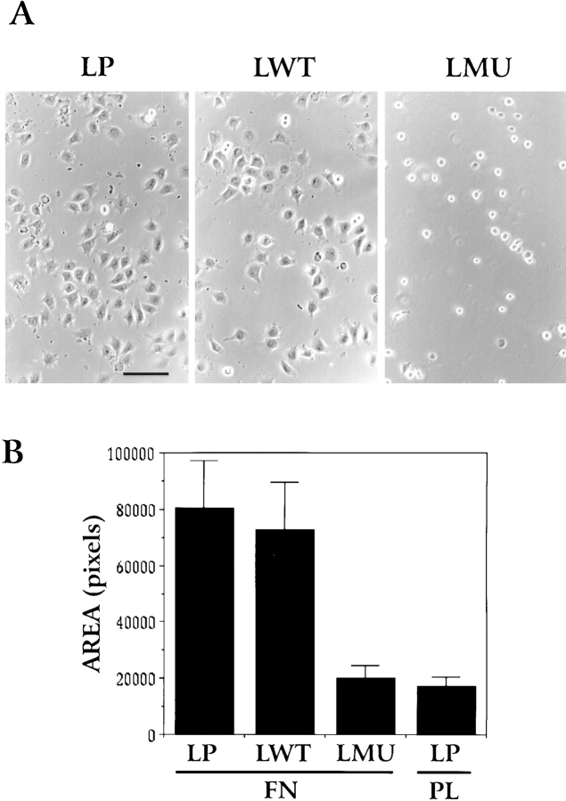
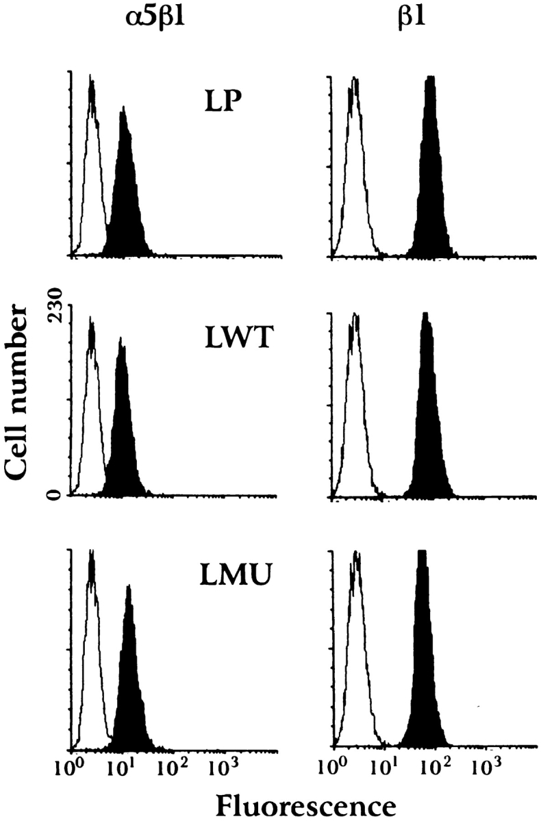
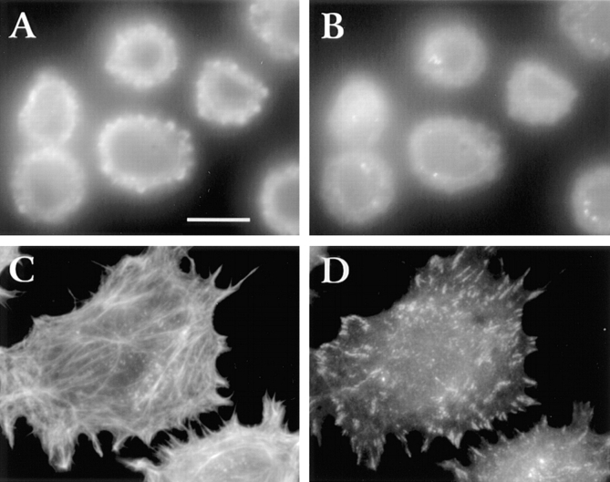
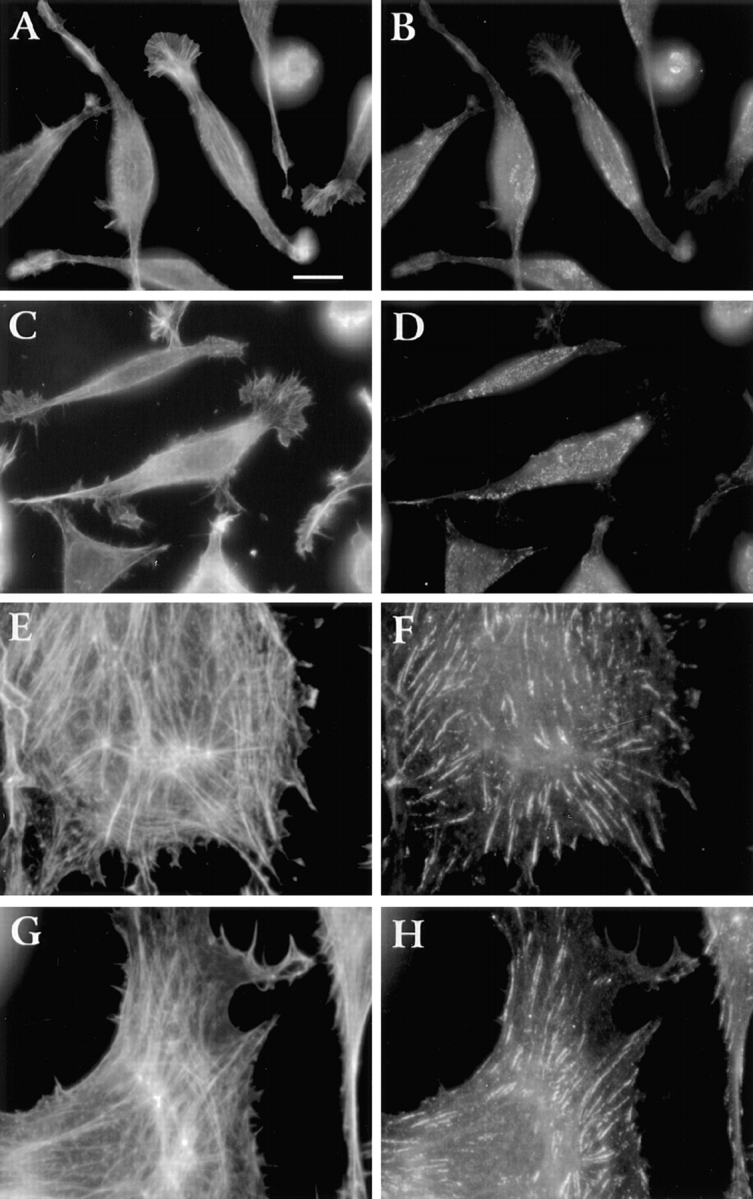
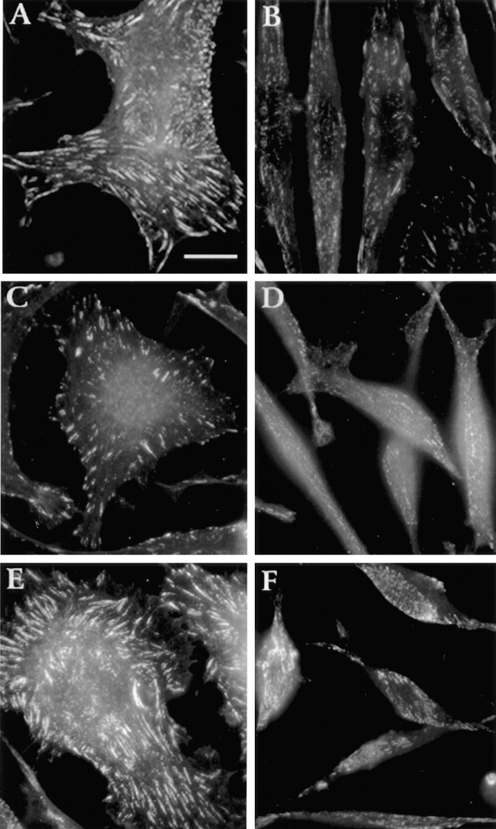
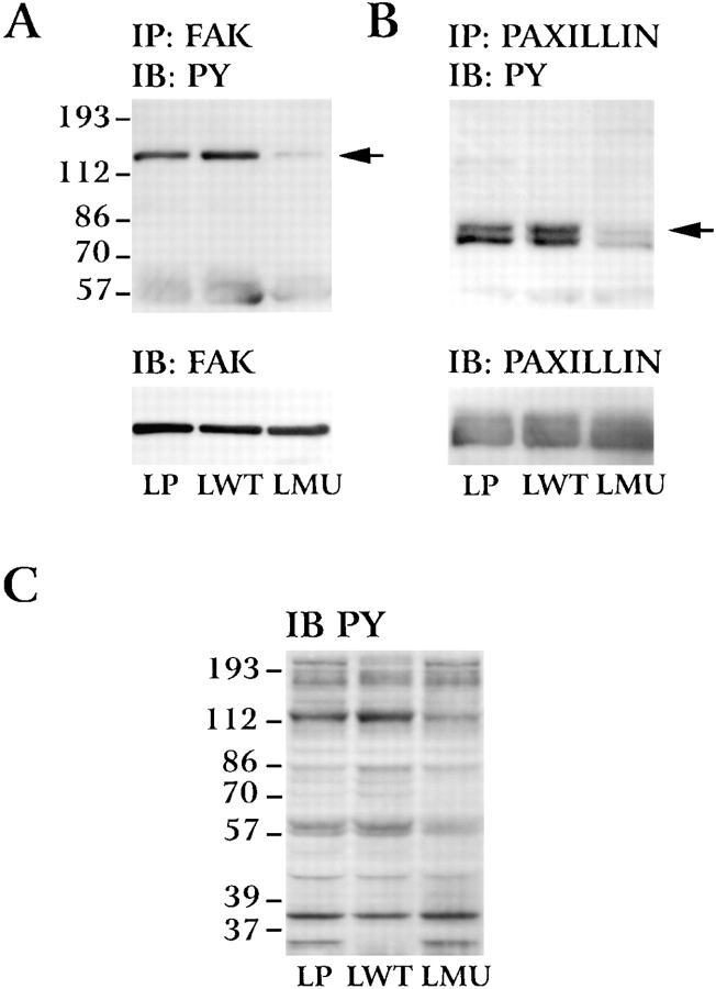
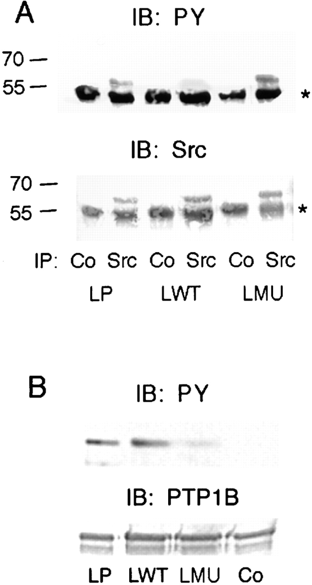
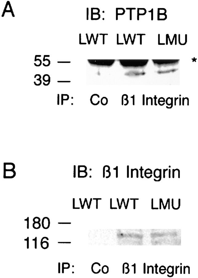
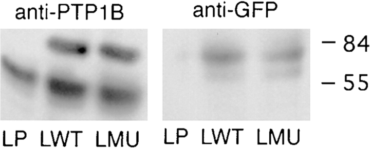
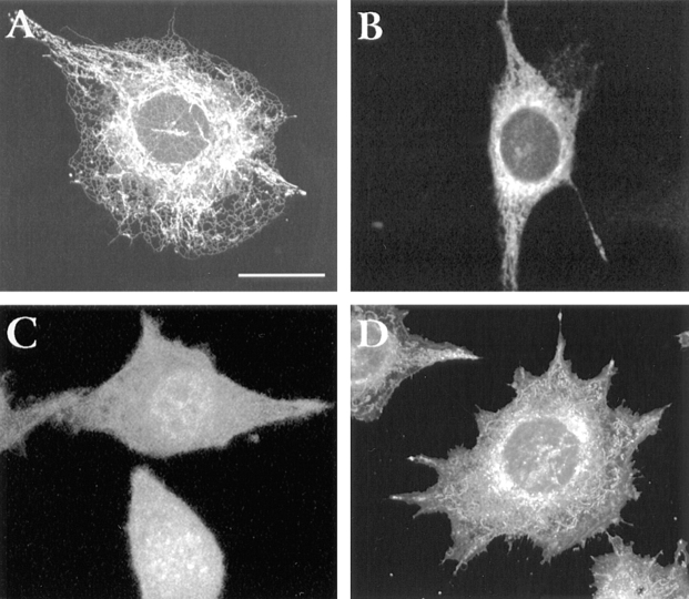
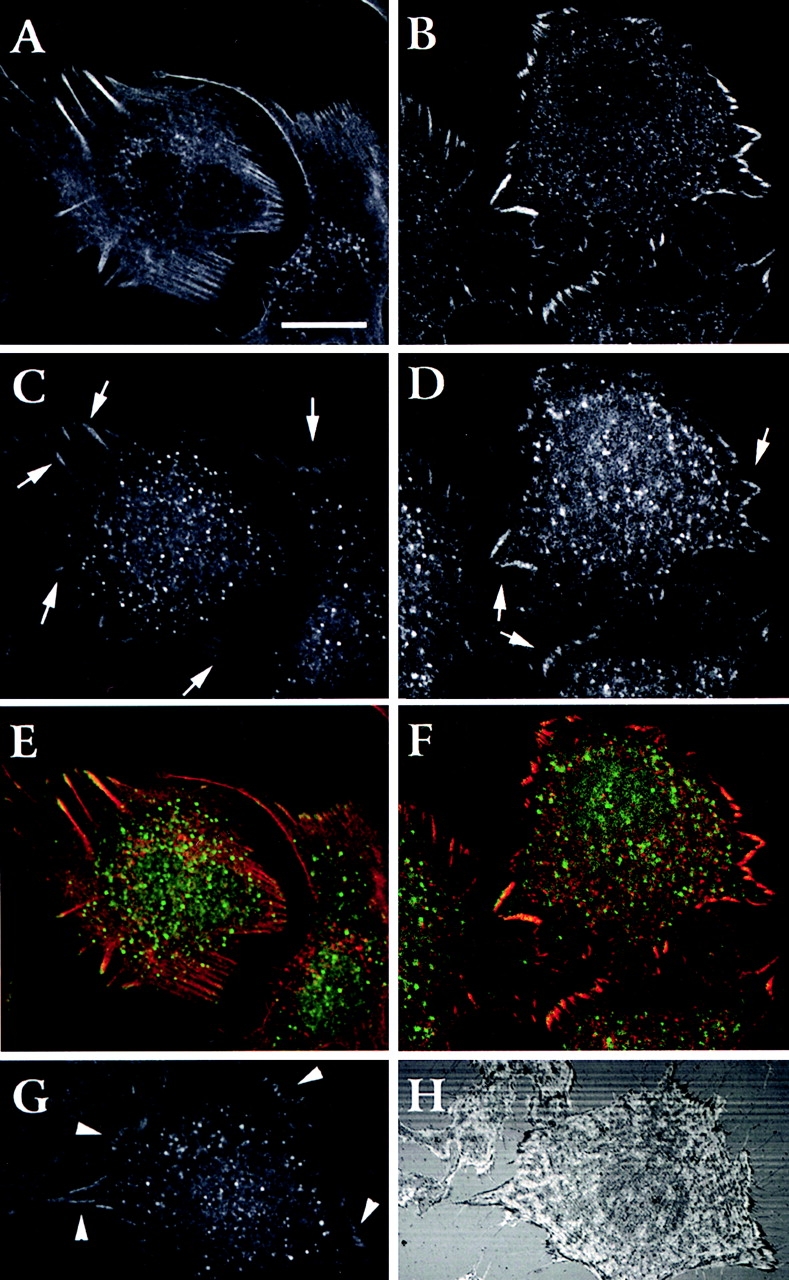
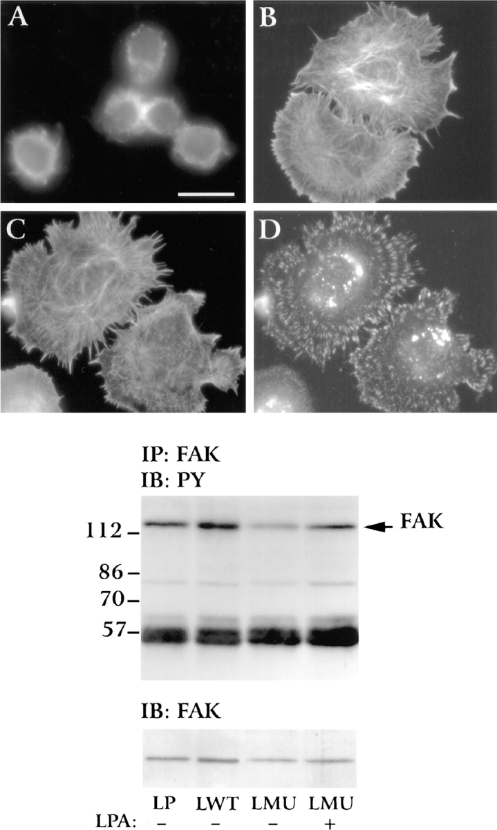
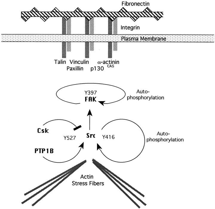
To investigate the role of nonreceptor protein tyrosine phosphatase 1B (PTP1B) in beta1-integrin- mediated adhesion and signaling, we transfected mouse L cells with normal and catalytically inactive forms of the phosphatase. Parental cells and cells expressing the wild-type or mutant PTP1B were assayed for (a) adhesion, (b) spreading, (c) presence of focal adhesions and stress fibers, and (d) tyrosine phosphorylation. Parental cells and cells expressing wild-type PTP1B show similar morphology, are able to attach and spread on fibronectin, and form focal adhesions and stress fibers. In contrast, cells expressing the inactive PTP1B have a spindle-shaped morphology, reduced adhesion and spreading on fibronectin, and almost a complete absence of focal adhesions and stress fibers. Attachment to fibronectin induces tyrosine phosphorylation of focal adhesion kinase (FAK) and paxillin in parental cells and cells transfected with the wild-type PTP1B, while in cells transfected with the mutant PTP1B, such induction is not observed. Additionally, in cells expressing the mutant PTP1B, tyrosine phosphorylation of Src is enhanced and activity is reduced. Lysophosphatidic acid temporarily reverses the effects of the mutant PTP1B, suggesting the existence of a signaling pathway triggering focal adhesion assembly that bypasses the need for active PTP1B. PTP1B coimmunoprecipitates with beta1-integrin from nonionic detergent extracts and colocalizes with vinculin and the ends of actin stress fibers in focal adhesions. Our data suggest that PTP1B is a critical regulatory component of integrin signaling pathways, which is essential for adhesion, spreading, and formation of focal adhesions.


Figures

Figure 1
Figure 1
(A) RT-PCR detection of chick PTP1B and β-actin mRNA expression in LP, LWT, or LMU cells. Shown is an ethidium bromide staining. (B) PTP1B protein expression. Equal amounts of protein in LP, LWT, or LMU cell homogenates were separated by SDS-PAGE and immunoblotted with anti-PTP1B antibody. Numbers to the right of the figure indicate the migration of prestained molecular mass standards in kilodaltons.
Figure 2
Figure 2
(A) Cell attachment of LP, LWT, and LMU cells to fibronectin. The number of cells attached after 45 min of plating was determined. Values for each condition represent the percentage of attached cells as compared with LP. Error bars are the standard deviation of three independent experiments, each counted in triplicate. Background attachment to BSA-coated wells was subtracted from the experimental values. In LP/RGD and LP/RAD, cells were preincubated 20 min on ice with the GRGDSP or GRADSP peptide (1 mM) and plated in the presence of the peptides. (B) Phase contrast images of LP, LWT, and LMU cells cultured in the presence of serum. Note the elongated and thin morphology of LMU cells compared with LWT and LP cells. Bar, 200 μm.
Figure 3
Figure 3
Spreading of LP, LWT, and LMU cells on fibronectin-coated coverslips. Cell spreading was determined after 30 min of plating. (A) Phase contrast images. (B) Cell area quantitated in pixels using the Metamorph image analysis system. Bar, 160 μm.
Figure 4
Figure 4
Flow cytometry analysis of the surface expression of α5β1-integrin fibronectin receptor and β1-integrin subunit in LP, LWT, and LMU cells. Cells were stained with either anti-α5β1 mAb, anti-β1 mAb (dark), or an isotype-matched control immunoglobulin (clear), followed by FITC-conjugated secondary antibody (see Materials and Methods).
Figure 5
Figure 5
Distribution of β1-integrin and actin in cells plated for 30 min on fibronectin. Serum-starved LMU (A and B) and LWT (C and D) cells in DME were plated on fibronectin-coated coverslips and fixed 30 min later. The cells were processed for double immunofluorescence staining with rat anti–mouse β1 mAb/FITC-goat anti–rat IgG (B and D) and TRITC-phalloidin (A and C). Note the lack of spreading, focal contacts, and actin stress fibers in LMU cells. Bar, 15 μm.
Figure 6
Figure 6
Distribution of β1-integrin and actin in cells cultured for 2 d. LMU (A–D), LWT (E and F), and LP (G and H) cells in DME/FBS were plated on fibronectin-coated coverslips. After 2 d, cells were fixed, permeabilized, and double stained with rat anti– β1 mAb/FITC-goat anti–rat IgG (B, D, F, and H) and TRITC-phalloidin (A, C, E, and G). Bar, 10 μm.
Figure 7
Figure 7
Distribution of FAK, paxillin, and vinculin in LMU and LWT cells plated on fibronectin-coated coverslips. LWT (A, C, and E) and LMU (B, D, and F) cells cultured for 2 d were fixed and processed for immunofluorescence using monoclonal antibodies against paxillin (A and B), FAK (C and D), or vinculin (E and F). Bar, 20 μm.
Figure 8
Figure 8
Phosphotyrosine levels of FAK and paxillin in LP, LWT, and LMU cells. LP, LWT, and LMU cells were plated on fibronectin for 30 min, lysed (see Materials and Methods), and immunoprecipitated (IP) with (A) anti-FAK mAb or (B) antipaxillin mAb and immunoblotted (IB) with antiphosphotyrosine (PY) antibody. Lower panels show stripped membranes reblotted with the same antibodies used for immunoprecipitation. (C) Phosphotyrosine staining of total proteins in Triton X-100 lysates. LP, LWT, and LMU cells plated on fibronectin for 30 min were lysed as described in Materials and Methods. Numbers at the left indicate the position of molecular mass markers in kilodaltons.
Figure 9
Figure 9
Comparison of phosphotyrosine levels and activity of Src in LP, LWT, and LMU cells. (A) Phosphotyrosine levels: serum-starved LP, LWT, and LMU cells were plated on fibronectin-coated plates and incubated for 1 h at 37°C. The cells were homogenized in RIPA buffer, immunoprecipitated with anti-Src antibody or control rabbit IgG (Co), and immunoblotted with antiphosphotyrosine mAb 4G10. The membrane was stripped and reblotted with anti-Src antibody. An asterisk to the right indicates the migration of IgG heavy chain. Numbers to the left indicate the position of molecular mass markers in kilodaltons. (B) Src activity: the anti-Src immunoprecipitates were assayed for kinase activity using recombinant, mutant, GST-chkPTP1B as substrate. The reaction mix was separated by SDS-PAGE, transferred to PVDF membranes, and immunoblotted with mAb 4G10. The same membranes were stripped and reblotted with anti-PTP1B antibody. Co, LP cells immunoprecipitated with control rabbit IgG.
Figure 10
Figure 10
PTP1B coimmunoprecipitation with β1-integrin. (A) LWT and LMU cells were lysed in nonionic detergent and immunoprecipitated with a rat anti–β1 antibody or normal rat IgG (Co). After separation by SDS-PAGE and transfer to PVDF membranes, the immunoprecipitates were reacted with a rabbit anti– PTP1B and developed with HRP-conjugated anti–rabbit antibody. (B) The membranes were stripped and reblotted with anti–β1-integrin antibody. An asterisk to the right indicates the migration of IgG heavy chain. Numbers at the left indicate the position of molecular mass markers in kilodaltons.
Figure 11
Figure 11
Expression of GFP–PTP1B. L cells were transfected with GFP– wtPTP1B, GFP–mutPTP1B, or GFP alone. The cells were homogenized in RIPA buffer, separated by SDS-PAGE, and immunoblotted with anti-PTP1B antibody or anti-GFP antibody. Numbers at the right indicate the position of molecular mass markers in kilodaltons.
Figure 12
Figure 12
Immunolocalization of GFP–wtPTP1B (A), GFP– mutPTP1B (B), and GFP alone (C) in L cells. L cells were transiently transfected and fixed 48 h later. For comparison, untransfected cells were stained with rhodamine B hexyl ester, a marker of the endoplasmic reticulum (D). Note that both wtPTP1B and mutPTP1B target predominantly to the ER. A single 1-μm optical section is shown for all fields. Bar, 30 μm.
Figure 13
Figure 13
Localization of wtPTP1B in focal adhesions. L cells were transiently transfected with GFP–wtPTP1B and fixed 48 h later. Cells stained with TRITC-phalloidin (A) or with a monoclonal antivinculin antibody followed by a rhodamine-anti–mouse antibody (B) were double stained with an anti-GFP polyclonal antibody followed by a FITC-anti–rabbit antibody (C and D). The distribution of the fluorescence in 1-μm-thick optical sections was examined by confocal microscopy. Arrows (C and D) show sites of overlapping staining at the tips of actin stress fibers and focal adhesions. Color images (E and F) show overlapping of the staining between actin-PTP1B (E) and vinculin-PTP1B (F). Orange and yellow indicates regions of overlapping. Anti-GFP staining (G) was compared with the pattern obtained by interference reflection microscopy (H). Arrowheads indicate the codistribution of the GFP– wtPTP1B with dark regions at the margins of the cell and where cell membrane form extensive focal adhesions. Bar, 25 μm.
Figure 14
Figure 14
Effect of lysophosphatidic acid in LMU cells. Serum-starved cells were plated in DME on fibronectin-coated coverslips in the absence or presence of 200 ng/ml lysophosphatidic acid and fixed after 15 min. Cells were permeabilized and stained with rat anti–β1 antibody (D) and TRITC-phalloidin labeled (A–C). Anti–β1-integrin staining was visualized by a FITC-conjugated goat anti–rat IgG. (A) LMU cells plated in the absence of LPA. (B) LWT cells plated in the absence of LPA. (C and D) LMU cells exposed to LPA and stained for actin (C) or β1-integrin (D). Note the formation of actin stress fiber (C) and induction of focal contacts (D) in LMU cells exposed to LPA. Tyrosine phosphorylation of FAK is shown in the immunoblot below. LP, LWT, and LMU cells were plated on fibronectin, lysed after 15 min under denaturing conditions, and immunoprecipitated with anti-FAK antibody. The immunoprecipitates were fractionated by SDS-PAGE and analyzed by immunoblotting with the 4G10 monoclonal antiphosphotyrosine antibody. Note the difference in the phosphorylation of FAK when LMU cells were plated in the absence or presence of LPA. The bottom panel shows the same blot stripped and probed with anti-FAK antibody. Numbers at the left indicate the position of molecular mass markers in kilodaltons. Bar, 20 μm.
Figure 15
Figure 15
Scheme showing a possible mechanism of action for PTP1B. Arrows indicate stimulatory interactions, and blunt lines indicate inhibitory interactions. The COOH-terminal Src tyrosine kinase, Csk, phosphorylates Y527-repressing Src activity. Activation of Src occurs by autophosphorylation of Y416 and dephosphorylation of Y527. Dephosphorylation of Y527 by PTP1B would therefore contribute to Src activation. Src, in turn, phosphorylates FAK and other components of the focal adhesion complex. The dominant-negative PTP1B would decrease dephosphorylation of Y527, maintaining Src in a repressed state.

Similar articles

Cited by

References

    1. Aberle H, Schwartz H, Kemler R. Cadherin-catenin complex: protein interactions and their implications for cadherin function. J Cell Biochem. 1996;61:514–523. - PubMed
    1. Balsamo J, Leung T-C, Ernst H, Zanin MKB, Hoffman S, Lilien J. Regulated binding of a PTP1B-like phosphatase to N-cadherin: control of cadherin-mediated adhesion by dephosphorylation of β-catenin. J Cell Biol. 1996;134:801–813. - PMC - PubMed
    1. Balsamo J, Arregui C, Leung TC, Lilien J. The nonreceptor protein tyrosine phosphatase PTP1B binds to the cytoplasmic domain of N-cadherin and regulates the cadherin-actin linkage. J Cell Biol. 1998;143:523–532. - PMC - PubMed
    1. Bandyopadhyay D, Kusari A, Kenner KA, Liu F, Chernoff J, Gustafson TA, Kusari J. Protein-tyrosine phosphatase 1B complexes with the insulin receptor in vivo and is tyrosine-phosphorylated in the presence of insulin. J Biol Chem. 1997;272:1639–1645. - PubMed
    1. Barry ST, Critchley DR. The rho-dependent assembly of focal adhesions in Swiss 3T3 cells is associated with increased tyrosine phosphorylation and the recruitment of both pp125FAK and protein kinase C-γ to focal adhesions. J Cell Sci. 1994;107:2033–2045. - PubMed

Publication types

MeSH terms