. 1998 Nov 16;188(10):1867-74.
doi: 10.1084/jem.188.10.1867.

Preselection thymocytes are more sensitive to T cell receptor stimulation than mature T cells

Affiliations

Preselection thymocytes are more sensitive to T cell receptor stimulation than mature T cells

G M Davey et al. J Exp Med. .

Abstract

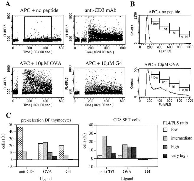
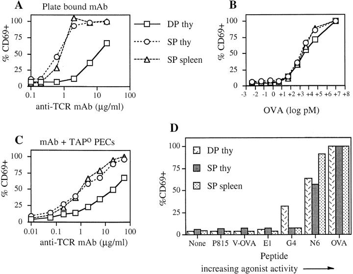
During T cell development, thymocytes which are tolerant to self-peptides but reactive to foreign peptides are selected. The current model for thymocyte selection proposes that self-peptide-major histocompatibility complex (MHC) complexes that bind the T cell receptor with low affinity will promote positive selection while those with high affinity will result in negative selection. Upon thymocyte maturation, such low affinity self-peptide-MHC ligands no longer provoke a response, but foreign peptides can incidentally be high affinity ligands and can therefore stimulate T cells. For this model to work, thymocytes must be more sensitive to ligand than mature T cells. Contrary to this expectation, several groups have shown that thymocytes are less responsive than mature T cells to anti-T cell receptor for antigen (TCR)/CD3 mAb stimulation. Additionally, the lower TCR levels on thymocytes, compared with T cells, would potentially correlate with decreased thymocyte sensitivity. Here we compared preselection thymocytes and mature T cells for early activation events in response to peptide-MHC ligands. Remarkably, the preselection thymocytes were more responsive than mature T cells when stimulated with low affinity peptide variants, while both populations responded equally well to the antigenic peptide. This directly demonstrates the increased sensitivity of thymocytes compared with T cells for TCR engagement by peptide-MHC complexes.


Figures

Figure 3
Figure 3
Changes in intracellular calcium in DP thymocytes and T cells stimulated with various ligands. (A) Dot plots of FL4/FL5 versus time for preselection (OT-I TAPo) DP thymocytes stimulated with APC alone, anti-CD3 mAb + goat anti–mouse Ig, OVAp + APC, and G4 + APC. The break in the trace corresponds to the time where the T cells and APCs were centrifuged together before being resuspended gently to pass through the flow cytometer. The two breaks for mAb cross-linking correspond to the sequential addition of anti-CD3 mAb and the goat anti–mouse Ig. (B) Representative histograms for FL4/FL5 of preselection DP thymocytes with APC alone or 10 μM OVAp, showing nonoverlapping markers set for low, intermediate, high, and very high expressing cells corresponding to increased intracellular calcium levels. Histogram data are based on the gate shown in A. (C) Graphs of the percentage of cells in each FL4/FL5 gate for preselection DP thymocytes and OT-I CD8 SP splenocytes stimulated with anti-CD3 mAb, OVAp, and G4 peptide. Markers were set as in B. Background levels determined by the APCs alone control have been subtracted from the values graphed. The APCs used in this experiment were of the 5AKb cell line. Similar results were seen using TAPo PEC as the APCs. The data shown are representative of four independent experiments.
Figure 1
Figure 1
Surface TCR levels are lower on DP thymocytes than T cells from OT-I mice. (A) Flow cytometry histograms of Vα2 TCR expression on splenic T cells (solid line) or DP thymocytes (dashed line) from OT-I mice and preselection DP thymocytes (dotted line) from OT-I TAPo mice. (B) Flow cytometry histograms of pan Vβ TCR expression on splenic T cells (solid line) or DP thymocytes (dashed line) from non-TCR Tg mice, splenic T cells (dash-dot line) from OT-I mice, and preselection DP thymocytes (dotted line) from OT-I TAPo mice.
Figure 1
Figure 1
Surface TCR levels are lower on DP thymocytes than T cells from OT-I mice. (A) Flow cytometry histograms of Vα2 TCR expression on splenic T cells (solid line) or DP thymocytes (dashed line) from OT-I mice and preselection DP thymocytes (dotted line) from OT-I TAPo mice. (B) Flow cytometry histograms of pan Vβ TCR expression on splenic T cells (solid line) or DP thymocytes (dashed line) from non-TCR Tg mice, splenic T cells (dash-dot line) from OT-I mice, and preselection DP thymocytes (dotted line) from OT-I TAPo mice.
Figure 2
Figure 2
CD69 upregulation on preselection DP thymocytes and T cells stimulated with various ligands. Immature and mature T cells were stimulated, for 3 h at 37°C, with dilutions of plate-bound anti-CD3 mAb (A), OVAp + APC (B), anti-CD3 + APC (C), or OVA variant peptides + APC (D). For preselection (OT-I TAPo) DP thymocytes (squares), OT-I CD8 SP thymocytes (circles), and OT-I CD8 SP splenocytes (triangles), the percentage of CD69-expressing cells on CD4/CD8-defined subpopulations was determined by flow cytometry. The data have been normalized such that the percentage of CD69+ cells in the presence of 5 μM OVAp = 100% for each cell subset. The graphs shown are representative of data from six to eight independent experiments.

References

    1. Jameson SC, Bevan MJ. T cell selection. Curr Opin Immunol. 1998;10:214–219. - PubMed
    1. Sebzda E, Wallace VA, Mayer J, Yeung RSM, Mak TW, Ohashi PS. Positive and negative thymocyte selection induced by different concentration of a single peptide. Science. 1994;263:1615–1618. - PubMed
    1. Ashton-Rickardt PG, Bandeira A, Delaney JR, Van Kaer L, Pircher H-P, Zinkernagel RM, Tonegawa S. Evidence for a differential avidity model of T cell selection in the thymus. Cell. 1994;76:651–663. - PubMed
    1. Hogquist KA, Jameson SC, Heath WR, Howard JL, Bevan MJ, Carbone FR. T cell receptor antagonist peptides induce positive selection. Cell. 1994;76:17–27. - PubMed
    1. Alam SM, Travers PJ, Wung JL, Nasholds W, Redpath S, Jameson SC, Gascoigne NRJ. T cell receptor affinity and thymocyte positive selection. Nature. 1996;381:616–620. - PubMed

Publication types