. 1998 Nov 16;188(10):1907-16.
doi: 10.1084/jem.188.10.1907.

Two enteropathogenic Escherichia coli type III secreted proteins, EspA and EspB, are virulence factors

Affiliations

Two enteropathogenic Escherichia coli type III secreted proteins, EspA and EspB, are virulence factors

A Abe et al. J Exp Med. .

Abstract

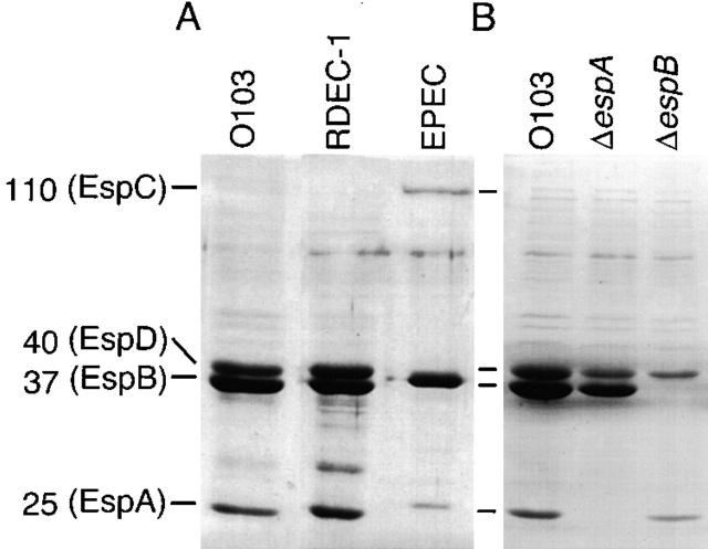
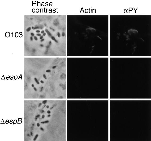
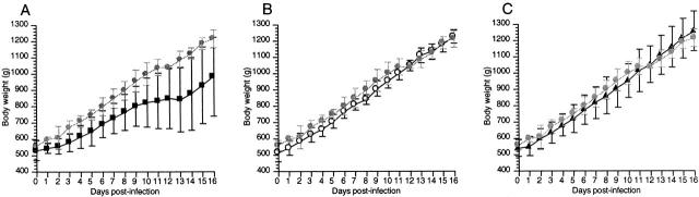
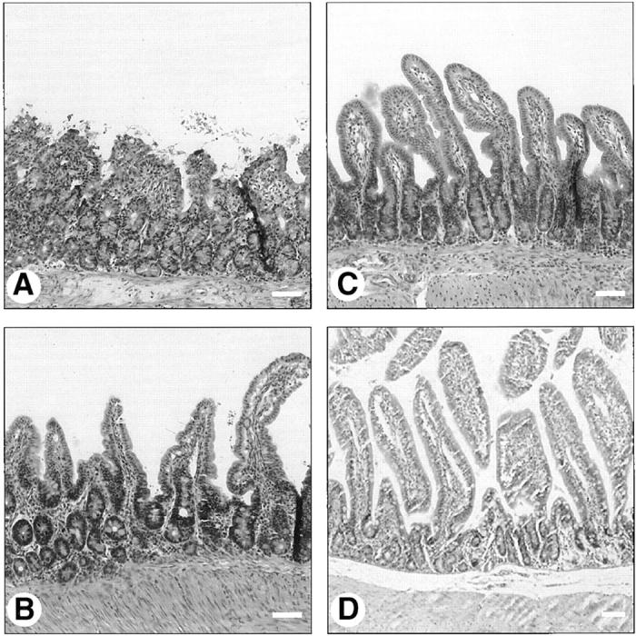
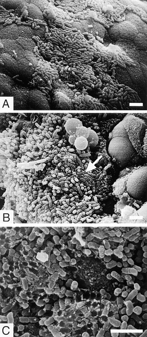
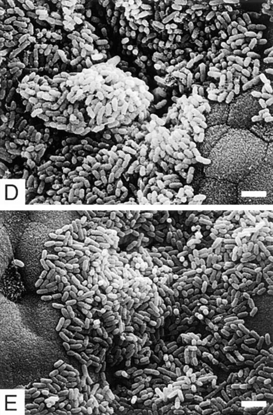
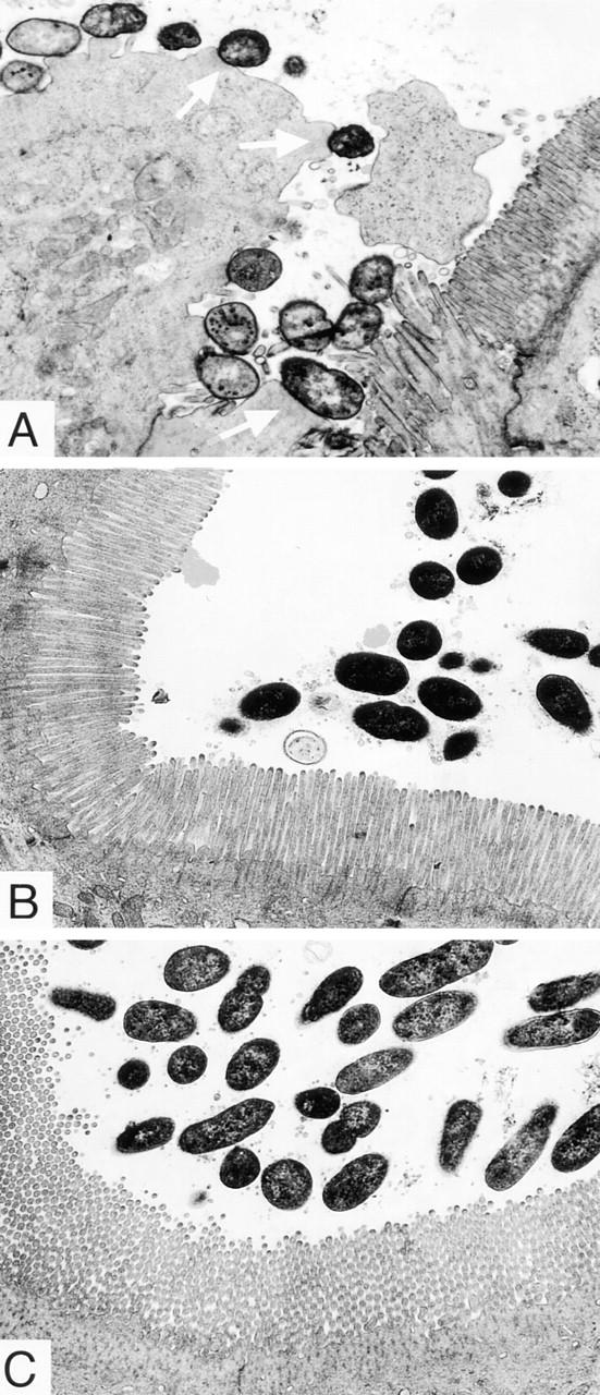
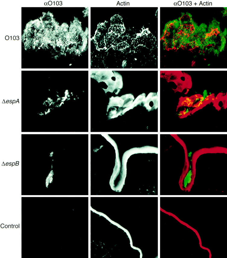
Enteropathogenic Escherichia coli (EPEC) belongs to a family of related bacterial pathogens, including enterohemorrhagic Escherichia coli (EHEC) O157:H7 and other human and animal diarrheagenic pathogens that form attaching and effacing (A/E) lesions on host epithelial surfaces. Bacterial secreted Esp proteins and a type III secretion system are conserved among these pathogens and trigger host cell signal transduction pathways and cytoskeletal rearrangements, and mediate intimate bacterial adherence to epithelial cell surfaces in vitro. However, their role in pathogenesis is still unclear. To investigate the role of Esp proteins in disease, mutations in espA and espB were constructed in rabbit EPEC serotype O103 and infection characteristics were compared to that of the wild-type strain using histology, scanning and transmission electron microscopy, and confocal laser scanning microscopy in a weaned rabbit infection model. The virulence of EspA and EspB mutant strains was severely attenuated. Additionally, neither mutant strain formed A/E lesions, nor did either one cause cytoskeletal actin rearrangements beneath the attached bacteria in the rabbit intestine. Collectively, this study shows for the first time that the type III secreted proteins EspA and EspB are needed to form A/E lesions in vivo and are indeed virulence factors. It also confirms the role of A/E lesions in disease processes.


Figures

Figure 1
Figure 1
Secreted protein profiles of REPEC O103, RDEC-1, EPEC (A), and REPEC O103 mutant strains (B). Bacteria were grown in DMEM and the secreted proteins were precipitated by addition of 10% TCA, and then analyzed by 12% SDS-PAGE stained with Coomassie blue. Strains: REPEC O103, O103; AAF004 (EspA), ΔespA; AAF005 (EspB), ΔespB.
Figure 2
Figure 2
Accumulation of cytoskeletal actin and tyrosine-phosphorylated proteins beneath bacteria in cultured epithelial cells. HeLa cells (105) were infected for 3 h with REPEC O103 or isogenic mutant strains. Infected HeLa cells were fixed by the addition of paraformaldehyde, then phalloidin-TxR and antiphosphotyrosine antibodies (αPY) were used for detection of colocalization of actin and tyrosine-phosphorylated proteins by immunofluorescence microscopy. Strains: REPEC O103, O103; AAF004 (EspA), ΔespA; AAF005 (EspB), ΔespB.
Figure 3
Figure 3
Growth curves of rabbits that survived infection with REPEC O103 (A) or those infected with AAF004 (B), and AAF005 (C). Bacteria (7 × 10⁸) were inoculated via the orogastric route and weights were measured daily. Values represent the average rabbit weight ± SD. Strains and control are indicated as follows: REPEC O103, black square; AAF004 (EspA), white circle; AAF005 (EspB), black triangle; PBS inoculation, gray circle.
Figure 4
Figure 4
Hematoxylin and eosin–stained ileal sections of rabbits infected with REPEC O103 (A), AAF004 (B), AAF005 (C), and PBS inoculation (D). Note the extensive villous blunting, epithelial cell necrosis and sloughing, and inflammation within the lamina propria of the wild-type infected tissue (A). In contrast, villous architecture and extent of inflammation within the lamina propria infected with AAF004 (EspA) or AAF005 (EspB) are similar to those of control animals that were inoculated PBS. Bar, 100 μm.
Figure 5
Figure 5
Scanning electron micrographs of the ileal villi infected with REPEC O103 (A–C), AAF004 (D), and AAF005 (E). REPEC O103, AAF004 (EspA), and AAF005 (EspB) adhered to the villi, and the adherence pattern is similar between wild-type and mutant strains. However, embedded bacteria as indicated by an arrow (B) and cup-like structures (C) were observed only in rabbits infected with REPEC O103. Ileal sections were taken 72 h (for REPEC O103 and AAF005) and 96 h (for AAF004) after infection. Bar, 4 μm.
Figure 5
Figure 5
Scanning electron micrographs of the ileal villi infected with REPEC O103 (A–C), AAF004 (D), and AAF005 (E). REPEC O103, AAF004 (EspA), and AAF005 (EspB) adhered to the villi, and the adherence pattern is similar between wild-type and mutant strains. However, embedded bacteria as indicated by an arrow (B) and cup-like structures (C) were observed only in rabbits infected with REPEC O103. Ileal sections were taken 72 h (for REPEC O103 and AAF005) and 96 h (for AAF004) after infection. Bar, 4 μm.
Figure 6
Figure 6
Transmission micrographs of the ileal villi infected with REPEC O103 (A), AAF004 (B), and AAF005 (C). The REPEC O103 are intimately associated with the ileal villi and form pedestal-like structures, which are indicated by arrows. Microbodies and elongated and swollen microvilli were also observed. In contrast, AAF004 (EspA) and AAF005 (EspB) do not cause A/E lesions, and intracellular damage was not seen. All ileal sections were taken 96 h after infection. Original magnification, ×6,300.
Figure 7
Figure 7
Confocal laser scanning micrographs of Peyer's patches infected with REPEC O103, AAF004, and AAF005. Peyer's patches were taken 72 h after infection and cryosections (20-μm-thick) were stained with phalloidin-TxR (red for overlay) and anti-O103 antibody (green for overlay). REPEC O103 adhered and colonized to the Peyer's patches, cytoskeletal actin beneath the attached bacteria was rearranged, and cup-like structures were also observed. In contrast, a small number of EspA and EspB mutant strains were observed in villi crypts, but no actin rearrangements occurred. Strains: REPEC O103, O103; AAF004 (EspA), ΔespA; AAF005 (EspB), ΔespB; PBS inoculation, Control.

References

    1. Donnenberg MS, Kaper JB. Enteropathogenic Escherichia coli. . Infect Immun. 1992;60:3953–3961. - PMC - PubMed
    1. Moon HW, Whipp SC, Argenzio RA, Levine MM, Giannella RA. Attaching and effacing activities of rabbit and human enteropathogenic Escherichia coliin pig and rabbit intestines. Infect Immun. 1983;41:1340–1351. - PMC - PubMed
    1. Francis DH, Collins JE, Duimstra JR. Infection of gnotobiotic pigs with an Escherichia coliO157:H7 strain associated with an outbreak of hemorrhagic colitis. Infect Immun. 1986;51:953–956. - PMC - PubMed
    1. Tzipori S, Wachsmuth KI, Smithers J, Jackson C. Studies in gnotobiotic piglets on non-0157:H7 Escherichia coliserotypes isolated from patients with hemorrhagic colitis. Gastroenterology. 1988;94:590–597. - PubMed
    1. Tzipori S, Karch H, Wachsmuth KI, Robins-Browne RM, O'Brien AD, Lior H, Cohen ML, Smithers J, Levine MM. Role of a 60-megadalton plasmid and Shiga-like toxins in the pathogenesis of infection caused by enterohemorrhagic Escherichia coliO157:H7 in gnotobiotic piglets. Infect Immun. 1987;55:3117–3125. - PMC - PubMed

Publication types

MeSH terms