. 1998 Nov 16;188(10):1929-39.
doi: 10.1084/jem.188.10.1929.

Oxazolone colitis: A murine model of T helper cell type 2 colitis treatable with antibodies to interleukin 4

Affiliations

Oxazolone colitis: A murine model of T helper cell type 2 colitis treatable with antibodies to interleukin 4

M Boirivant et al. J Exp Med. .

Abstract

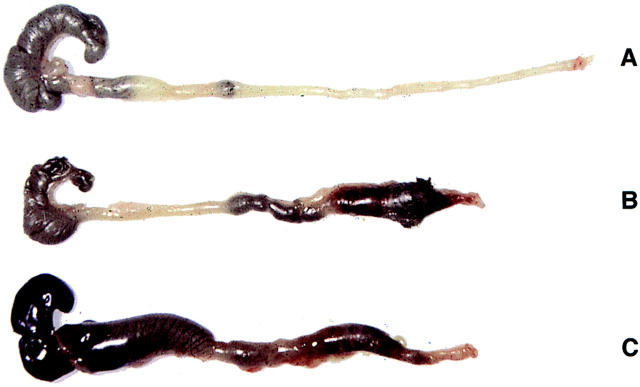
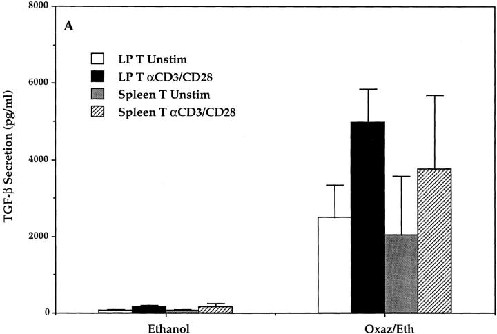
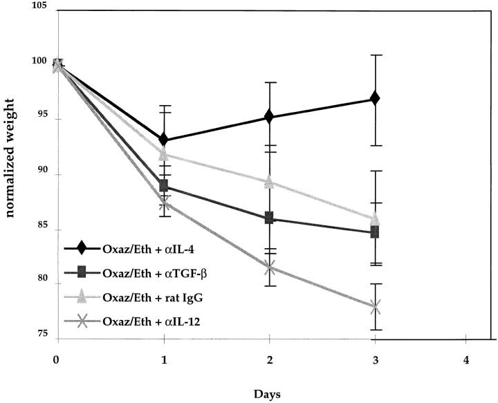
In this study we describe oxazolone colitis, a new form of experimental colitis. This model is induced in SJL/J mice by the rectal instillation of the haptenating agent, oxazolone, and is characterized by a rapidly developing colitis confined to the distal half of the colon; it consists of a mixed neutrophil/lymphocyte infiltration limited to the superficial layer of the mucosa which is associated with ulceration. Oxazolone colitis is a T helper cell type 2 (Th2)-mediated process since stimulated T cells from lesional tissue produce markedly increased amounts of interleukin (IL)-4 and IL-5; in addition, anti-IL-4 administration leads to a striking amelioration of disease, whereas anti-IL-12 administration either has no effect or exacerbates disease. Finally, this proinflammatory Th2 cytokine response is counterbalanced by a massive transforming growth factor-beta (TGF-beta) response which limits both the extent and duration of disease: lesional (distal) T cells manifest a 20-30-fold increase in TGF-beta production, whereas nonlesional (proximal) T cells manifest an even greater 40-50-fold increase. In addition, anti-TGF-beta administration leads to more severe inflammation which now involves the entire colon. The histologic features and distribution of oxazolone colitis have characteristics that resemble ulcerative colitis (UC) and thus sharply distinguish this model from most other models, which usually resemble Crohn's disease. This feature of oxazolone colitis as well as its cytokine profile have important implications to the pathogenesis and treatment of UC.


Figures

Figure 1
Figure 1
Wasting disease in mice with oxazolone colitis. Intrarectal administration of oxazolone induces rapid onset of severe bloody diarrhea and wasting disease. Shown are weight changes over a 10-d period occurring in normal SJL/J control mice treated with 50% ethanol alone, and SJL/J mice treated with oxazolone in 50% ethanol. Data are from one representative experiment (out of a total of three experiments). Each point represents average weight data pooled from five mice. Standard errors are indicated.
Figure 2
Figure 2
Macroscopic changes of colons in hapten-treated mice. Photographs of dissected large intestine of (A) normal SJL/J control mouse treated with 50% ethanol, (B) SJL/J mouse treated with oxazolone in 50% ethanol 2 d after initial rectal administration, and (C) appearance of colons of mice with TNBS-induced colitis 7 d after intrarectal administration. The colons of oxazolone-treated mice display a hemorrhagic edematous colon limited to the distal half of the colon. This is in contrast to colons in TNBS-treated mice which display an inflammation of the entire colon.
Figure 3
Figure 3
Histologic analysis of the colons from SJL/J mice with hapten-induced colitis and control mice. (A) Photomicrograph of hematoxylin and eosin– stained (H/E) paraffin section of distal colon (×100) from an oxazolone-treated mouse on day 2. Significant edema with inflammatory infiltrates localized to the superficial mucosal layer is present. (B) H/E-stained section of colon (×100) from a TNBS-treated mouse at 7 d after intrarectal administration: a severe transmural colitis with bowel wall thickening is seen. (C) Photomicrograph of H/E-stained section of colon (×150) from an oxazolone-treated mouse showing the presence of superficial hemorrhage, ulceration, distortion of the crypts, loss of goblet cells, and mucin depletion. (D) High-power micrograph of cross-section of H/E-stained section of colon (×400) of an oxazolone-treated mouse, showing a mixed lymphocytic infiltrate localized to the superficial layers of the mucosa and the presence of a luminal cellular exudate. (E) Photomicrograph of H/E-stained cross-section from the proximal colon (×100) of an oxazolone-treated mouse showing only a small amount of mucosal edema and lymphocytic infiltrate. (F) Photomicrograph of H/E-stained cross-section of normal control (×100) colon 2 d after ethanol administration.
Figure 4
Figure 4
Cytokine production of stimulated and unstimulated LP and spleen T cells in oxazolone-induced colitis. LP T and spleen T cells were isolated from oxazolone and control (ethanol-treated) mice on day 2 and cultured for 48 h in the presence or absence of anti-CD3 and anti-CD28 (see Materials and Methods). Culture supernatants were analyzed for concentrations of IL-4, IL-5, and IFN-γ secretion by specific ELISA. Data shown represent pooled values from three independent experiments. Standard errors are indicated.
Figure 4
Figure 4
Cytokine production of stimulated and unstimulated LP and spleen T cells in oxazolone-induced colitis. LP T and spleen T cells were isolated from oxazolone and control (ethanol-treated) mice on day 2 and cultured for 48 h in the presence or absence of anti-CD3 and anti-CD28 (see Materials and Methods). Culture supernatants were analyzed for concentrations of IL-4, IL-5, and IFN-γ secretion by specific ELISA. Data shown represent pooled values from three independent experiments. Standard errors are indicated.
Figure 5
Figure 5
In vitro IL-4 secretion by LP T cells extracted from proximal (noninvolved) and distal (involved) colons of mice with oxazolone- induced colitis as compared with control (ethanol-treated) mice. Data shown represent pooled values from three independent experiments. Standard errors are indicated.
Figure 6
Figure 6
(A) TGF-β secretion of unstimulated and stimulated LP and spleen T cells (LP T cells isolated from whole colons of mice with oxazolone colitis). LP T and spleen T cells were isolated from oxazolone and control (ethanol-treated) mice on day 2, cultured for 48 h in the presence or absence of anti-CD3 and anti-CD28, and culture supernatants were analyzed for concentration of TGF-β secretion by specific ELISA. Data shown represent pooled values from three independent experiments. Standard errors are indicated. (B) In parallel experiments, purified LP T cells were extracted from proximal (noninvolved) and distal (involved) colons of mice with oxazolone colitis and compared with control (ethanol-treated) mice for TGF-β secretion. Data shown represent pooled values from three independent experiments. Standard errors are indicated.
Figure 6
Figure 6
(A) TGF-β secretion of unstimulated and stimulated LP and spleen T cells (LP T cells isolated from whole colons of mice with oxazolone colitis). LP T and spleen T cells were isolated from oxazolone and control (ethanol-treated) mice on day 2, cultured for 48 h in the presence or absence of anti-CD3 and anti-CD28, and culture supernatants were analyzed for concentration of TGF-β secretion by specific ELISA. Data shown represent pooled values from three independent experiments. Standard errors are indicated. (B) In parallel experiments, purified LP T cells were extracted from proximal (noninvolved) and distal (involved) colons of mice with oxazolone colitis and compared with control (ethanol-treated) mice for TGF-β secretion. Data shown represent pooled values from three independent experiments. Standard errors are indicated.
Figure 7
Figure 7
Cytokine production by LP T cells from resolving oxazolone colitis. LP T cells isolated from the distal colons of mice with oxazolone colitis and control ethanol-treated mice at 10 d after intrarectal administration were stimulated in vitro as indicated in the legend to Fig. 4. Culture supernatants were assayed for IL-4, IL-5, and TGF-β by specific ELISA. Data shown represent pooled values from three independent experiments. Standard errors are indicated.
Figure 7
Figure 7
Cytokine production by LP T cells from resolving oxazolone colitis. LP T cells isolated from the distal colons of mice with oxazolone colitis and control ethanol-treated mice at 10 d after intrarectal administration were stimulated in vitro as indicated in the legend to Fig. 4. Culture supernatants were assayed for IL-4, IL-5, and TGF-β by specific ELISA. Data shown represent pooled values from three independent experiments. Standard errors are indicated.
Figure 8
Figure 8
Anti–IL-4 antibodies can prevent the onset of oxazolone colitis. Weight changes in SJL/J mice who received intrarectal oxazolone administration and then treated with either rat IgG control Ab, anti– TGF-β mAb, or anti–IL-12 mAb. After initial reduction in body weight in all groups at day 1, the mice treated with anti–IL-4 showed a significant increase in body weight, whereas mice treated with either IgG control Ab, anti–TGF-β, or anti–IL-12 continued to lose body weight. Each point represents pooled data from five mice. Standard errors are indicated.
Figure 9
Figure 9
Macroscopic appearance of colons obtained from (A) control (ethanol-treated) mice; SJL/J mice who received intrarectal oxazolone or intrarectal oxazolone plus (B) IgG control Ab; (C) anti–IL-4; (D) anti– TGF-β mAb; or (E) anti–IL-12 mAb (all antibodies were administered intraperitoneally). Mice treated with anti–IL-4 mAb showed a significant resolution in inflammation, whereas mice treated with anti–TGF-β reveal a severe colitis that now involves the entire colon. Colons from one representative experiment out of two are shown.
Figure 10
Figure 10
Cytokine production of stimulated and unstimulated LP T cells in oxazolone-induced colitis mice treated with rat IgG control Ab, anti–IL-4, anti–TGF-β mAb, or anti–IL-12 mAb at the time of intrarectal oxazolone administration. LP T were isolated on day 3, cultured for 48 h in the presence or absence of anti-CD3 and anti-CD28, and culture supernatants were analyzed for concentration of (A) IL-4 and (B) TGF-β by specific ELISA. Data from one representative experiment out of two are shown.
Figure 10
Figure 10
Cytokine production of stimulated and unstimulated LP T cells in oxazolone-induced colitis mice treated with rat IgG control Ab, anti–IL-4, anti–TGF-β mAb, or anti–IL-12 mAb at the time of intrarectal oxazolone administration. LP T were isolated on day 3, cultured for 48 h in the presence or absence of anti-CD3 and anti-CD28, and culture supernatants were analyzed for concentration of (A) IL-4 and (B) TGF-β by specific ELISA. Data from one representative experiment out of two are shown.

References

    1. Neurath MF, Fuss I, Kelsall BL, Stüber E, Strober W. Antibodies to IL-12 abrogate established experimental colitis in mice. J Exp Med. 1995;182:1281–1290. - PMC - PubMed
    1. Fuss IJ, Neurath MF, Boirivant M, Klein JS, de la Motte C, Strong SA, Fiocchi C, Strober W. Disparate CD4+lamina propria (LP) lymphokine secretion profiles in inflammatory bowel disease. Crohn's disease LP cells manifest increased secretion of IFN-γ, whereas ulcerative colitis LP cells manifest increased secretion of IL-5. J Immunol. 1996;157:1261–1270. - PubMed
    1. Parronchi P, Romagni P, Annunziato F, Sampognaro S, Becchio A, Giannarini L, Maggi E, Pupilli C, Tonelli F, Romagni S. Type 1 T-helper cell predominance and IL-12 expression in the gut of patients with Crohn's disease. Am J Pathol. 1997;150:823–832. - PMC - PubMed
    1. Breese E, Braegger CP, Corrigan CJ, Walker-Smith JA, MacDonald TT. Interleukin-2 and interferon-γ secreting T cells in normal and diseased human intestinal mucosa. Immunology. 1993;78:127–131. - PMC - PubMed
    1. Monteleone G, Biancone L, Marasco R, Marrone G, Marasco O, Luzza F, Pallone F. Interleukin-12 is expressed and actively released by Crohn's disease intestinal lamina propria mononuclear cells. Gastroenterology. 1997;112:1169–1178. - PubMed