. 1998 Nov 16;188(10):1941-53.
doi: 10.1084/jem.188.10.1941.

Development of a natural model of cutaneous leishmaniasis: powerful effects of vector saliva and saliva preexposure on the long-term outcome of Leishmania major infection in the mouse ear dermis

Affiliations

Development of a natural model of cutaneous leishmaniasis: powerful effects of vector saliva and saliva preexposure on the long-term outcome of Leishmania major infection in the mouse ear dermis

Y Belkaid et al. J Exp Med. .

Abstract

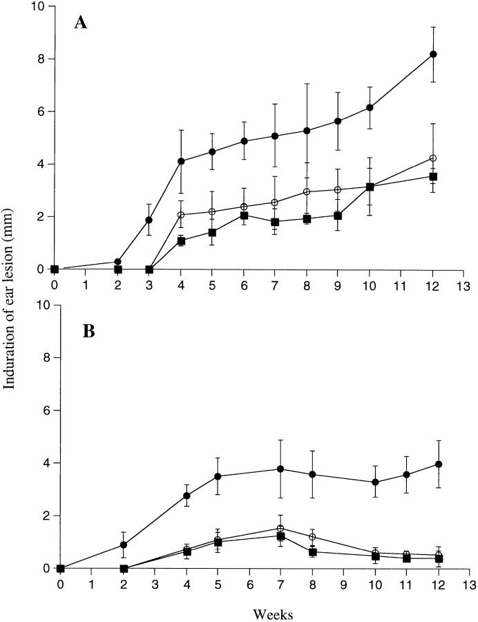
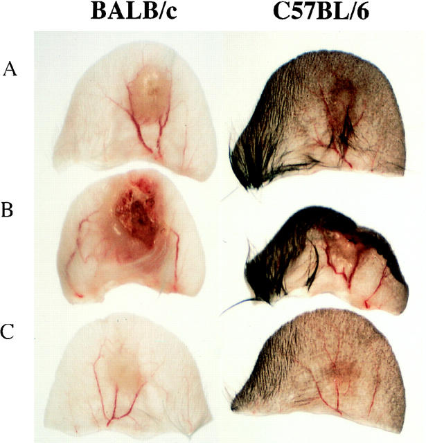
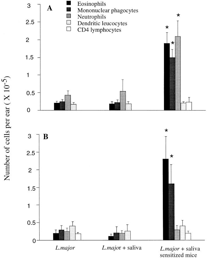
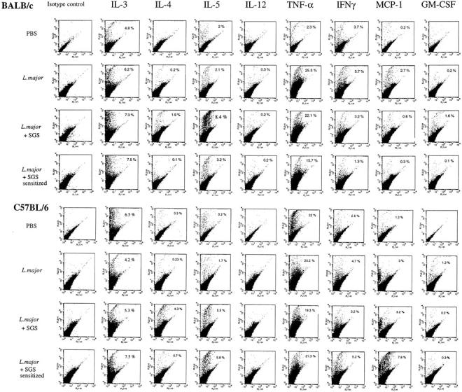
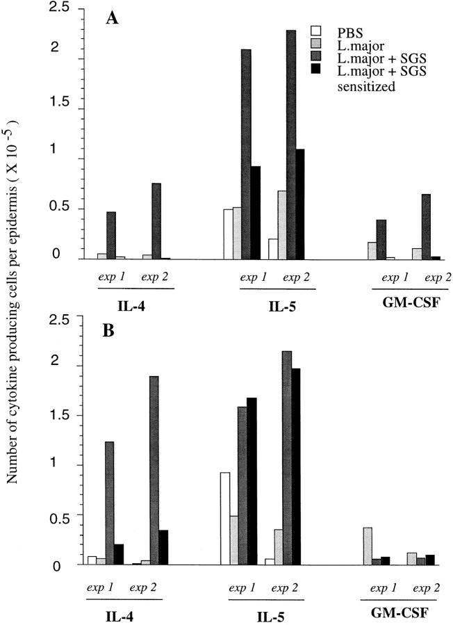
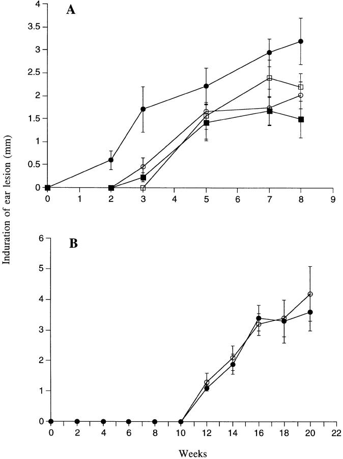
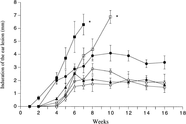
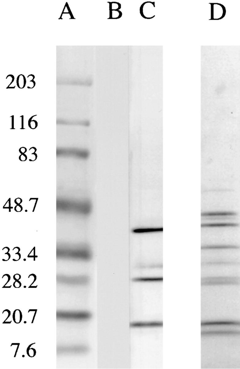
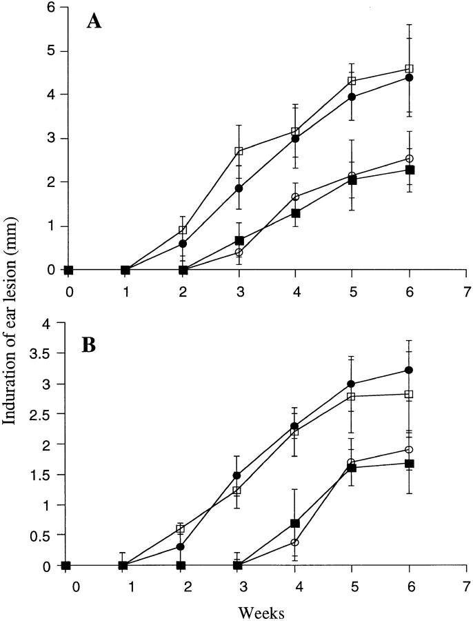
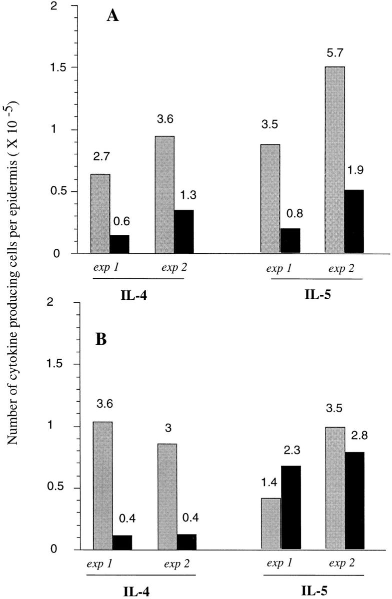
We have developed a model of cutaneous leishmaniasis due to Leishmania major that seeks to mimic the natural conditions of infection. 1,000 metacyclic promastigotes were coinoculated with a salivary gland sonicate (SGS) obtained from a natural vector, Phlebotomus papatasii, into the ear dermis of naive mice or of mice preexposed to SGS. The studies reveal a dramatic exacerbating effect of SGS on lesion development in the dermal site, and a complete abrogation of this effect in mice preexposed to salivary components. In both BALB/c and C57Bl/6 (B/6) mice, the dermal lesions appeared earlier, were more destructive, and contained greater numbers of parasites after infection in the presence of SGS. Furthermore, coinoculation of SGS converted B/6 mice into a nonhealing phenotype. No effect of SGS was seen in either IL-4- deficient or in SCID mice. Disease exacerbation in both BALB/c and B/6 mice was associated with an early (6 h) increase in the frequency of epidermal cells producing type 2 cytokines. SGS did not elicit type 2 cytokines in the epidermis of mice previously injected with SGS. These mice made antisaliva antibodies that were able to neutralize the ability of SGS to enhance infection and to elicit IL-4 and IL-5 responses in the epidermis. These results are the first to suggest that for individuals at risk of vector-borne infections, history of exposure to vector saliva might influence the outcome of exposure to transmitted parasites.


Figures

Figure 1
Figure 1
Size of induration of the ear lesion for (A) BALB/c or (B) C57BL/6 mice after intradermal inoculation of 1,000 L. major metacyclic promastigotes alone (○) or with 0.2 pair of SGS to naive mice (•) or to SGS presensitized mice (▪). Mean induration in mm ± 1 SD, 8–10 mice per group.
Figure 2
Figure 2
Photomicrograph of BALB/c or C57BL/6 ears 10 wk after intradermal inoculation of 1,000 L. major metacyclic promastigotes alone (A), or with 0.2 pair of SGS to naive (B), or to SGS presensitized mice (C).
Figure 3
Figure 3
Leukocytes present in the dermal compartment after intradermal inoculation of L. major into the ear of (A) BALB/c or (B) C57BL/6 mice. Mice were injected with 1,000 of L. major metacyclic promastigotes alone or with 0.2 pair of SGS to naive or to SGS presensitized mice. The ears were processed 18 h after inoculation, the cells obtained from four to five ears per group were pooled, and the different populations of leukocytes were identified by staining and flow cytometry as described in Materials and Methods. The data shown are the mean ± 1 SD of three separate experiments. * Values significantly greater (P < 0.001) than the values in the comparable cell populations of the two other groups.
Figure 4
Figure 4
Two-parameter dot plots of epidermal cells of BALB/c and C57BL/6 mice stained for intracellular cytokines produced at 6 h in response to the intradermal inoculation of PBS, or 1,000 L. major metacyclic promastigotes alone, or 1,000 metacyclics plus 0.2 pair of SGS, to naive or to SGS presensitized mice. After recovery, the epidermal cells from six ears per group, were pooled and preincubated with Brefeldin A for 6 h before being stained. Numbers represent the percentage of cells with FL-2 signals for the particular cytokine that were greater than the background signals established using the PE-isotype control (IgG1). The data shown are from a single experiment, representative of two separate experiments.
Figure 5
Figure 5
Total number of epidermal cells per ear producing type 2 cytokines in (A) BALB/c and (B) C57Bl/6 mice, 6 h after intradermal inoculation of PBS, L. major, L. major + SGS, and L. major + SGS in SGS-sensitized mice. Data from two separate experiments are shown, determined from the two-color FACS® analysis of epidermal cells pooled from 6 ears per group, and calculated as the average number of epidermal cells per ear × the percentage of cells staining positive for each cytokine.
Figure 6
Figure 6
Size of the induration of the ear lesion in (A) BALB/c wild-type mice (○, •) or in IL-4–deficient BALB/c mice (□, ▪) after intradermal inoculation of 1,000 metacyclic promastigotes alone (open symbols) or with 0.2 pair of SGS (closed symbols) and in (B) BALB/c SCID mice inoculated with the parasite alone (○) or with 0.2 pair of SGS (•). Mean induration in mm ± 1 SD, six to eight mice per group.
Figure 7
Figure 7
Size of the induration of the ear lesion in C57Bl/6 wild-type mice (○, •); C57Bl/6 IL-4–deficient mice (▵, ▴); or C57Bl/6 IL-12p40–deficient mice (□, ▪) after intradermal inoculation of 1,000 metacyclic promastigotes alone (open symbols) or with 0.2 pair of SGS (closed symbols). Mean induration in mm ± 1 SD, 6–10 mice per group.
Figure 8
Figure 8
Western blot analysis of salivary glands (1 gland per lane) using pooled sera from unimmunized BALB/c mice (lane B), BALB/c mice injected intradermally two times with 0.2 pair SGS (lane C); Coomassie blue– stained gel before transfer is shown in lane D; molecular weight markers are shown in lane A.
Figure 9
Figure 9
Size of the induration of the ear lesion in (A) BALB/c and (B) C57BL/6 mice after intradermal inoculation of 1,000 metacyclic promastigotes alone (○) or with 0.2 pair of SGS saliva (•), with 0.2 pair of SGS preincubated with serum IgG from normal mice (□), or with 0.2 pair of SGS preincubated with serum IgG from BALB/c or C57Bl/6 mice injected twice with SGS (▪). Mean induration in mm ± 1 SD, six mice per group.
Figure 10
Figure 10
Total number of epidermal cells per ear staining for selected cytokines in (A) BALB/c and (B) C57Bl/6 mice, 6 h after intradermal inoculation of 1,000 metacyclic promastigotes plus 0.2 gland SGS preincubated with control serum IgG (gray bars) or with serum IgG from SGS presensitized mice (solid bars). Data from two separate experiments are shown, determined from the two-color FACS® analysis of epidermal cells pooled from six ears per group, and calculated as the average number of epidermal cells per ear × the percentage of cells staining positive for each cytokine (numbers above each bar).

References

    1. Warburg A, Schlein Y. The effect of post-bloodmeal nutrition of Phlebotomus papatasii on the transmission of Leishmania major. . Am J Trop Med Hyg. 1986;35:926–930. - PubMed
    1. Ribeiro JMC. Role of saliva in blood-feeding by arthropods. Annu Rev Entomol. 1987;32:463–478. - PubMed
    1. Hall LR, Titus RG. Sand fly saliva selectively modulates macrophage functions that inhibit killing of Leishmania majorand nitric oxide production. J Immunol. 1995;155:3501–3506. - PubMed
    1. Titus RG. Salivary gland lysate from the Sand fly Lutzomia longipalpissuppresses the immune response of mice to sheep red blood cells in vivo and concanavalin A in vitro. Exp Parasitol. 1998;89:133–136. - PubMed
    1. Wikel, S.K. 1996. Immunology of the skin. In The Immunology of Host-Ectoparasitic Arthropod Relationships. S.K. Wikel, editor. CAB International, Wallingford, U.K. 1–29.

MeSH terms