. 1998 Nov 5;396(6706):92-6.
doi: 10.1038/23974.

Structure of the haemagglutinin-esterase-fusion glycoprotein of influenza C virus

Affiliations

Structure of the haemagglutinin-esterase-fusion glycoprotein of influenza C virus

P B Rosenthal et al. Nature. .

Abstract

The spike glycoproteins of the lipid-enveloped orthomyxoviruses and paramyxoviruses have three functions: to recognize the receptor on the cell surface, to mediate viral fusion with the cell membrane, and to destroy the receptor. In influenza C virus, a single glycoprotein, the haemagglutinin-esterase-fusion (HEF) protein, possesses all three functions. In influenza A and B, the first two activities are mediated by haemagglutinin and the third by a second glycoprotein, neuraminidase. Here we report the crystal structure of the HEF envelope glycoprotein of influenza C virus. We have identified the receptor-binding site and the receptor-destroying enzyme (9-O-acetylesterase) sites, by using receptor analogues. The receptor-binding domain is structurally similar to the sialic acid-binding domain of influenza A haemagglutinin, but binds 9-O-acetylsialic acid. The esterase domain has a structure similar to the esterase from Streptomyces scabies and a brain acetylhydrolase. The receptor domain is inserted into a surface loop of the esterase domain and the esterase domain is inserted into a surface loop of the stem. The stem domain is similar to that of influenza A haemagglutinin, except that the triple-stranded, alpha-helical bundle diverges at both of its ends, and the amino terminus of HEF2, the fusion peptide, is partially exposed. The segregation of HEF's three functions into structurally distinct domains suggests that the entire stem region, including sequences at the amino and carboxy termini of HEF1 which precede the post-translational cleavage site between HEF1 and HEF2, forms an independent fusion domain which is probably derived from an ancestral membrane fusion protein.


Figures

Figure 1
Figure 1. Haemagglutinin-esterase-fusion glycoprotein structure.
a, The structure of the HEF trimer. HEF1 (blue), HEF2 (red), receptor analogue and enzyme inhibitor ligands, yellow; N -linked carbohydrate ball-and-stick (purple). HEF1 is linked to HEF2 by a disulfide bond from Cys 6 of HEF1 to Cys 137 of HEF2. b, Monomer surface of HEF (Grasp) showing 9-O -acetylsialoside receptor binding site (top) and 9-O -acetylesterase site (bottom). Inset, the esterase removes the acetyl group of 9-O -acetylsialic acid (see arrow).
Figure 2
Figure 2. Comparison of HEF and HA monomers.
a, HEF monomer structure and exploded views of the sequence segments (coloured by domain). Superposition of corresponding HA (X:31 strain; PDB code 1hge) segments (grey) are shown with r.m.s. values below the segment name. Fusion peptides are yellow. b, Linear order of the sequence segments in HEF, coloured by domains. Red segments: F1, F2, F3; green: E1, E2; light green: E′; blue: R. c, Topological relationship of the compact domains in HEF. d, HA monomer structure with sequence segments coloured by domains. e, Linear order of the sequence segments in HA, coloured by domain. Red segments: F1, F2, F3; light green: E′; blue: R.
Figure 3
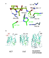
Figure 3. HEF receptor binding.
a, Ligand bound to the receptor-binding site. Potential hydrogen bonds are indicated in green or red for those conserved in HA ligand binding. Four polar contacts are formed with the ligand identically in HEF and HA: two from the hydroxyl group of HEF1 Tyr 127 (Y98 in HA1) to the 8-hydroxyl and 9-amide of the ligands and two from main-chain atoms: the carbonyl oxygen of HEF1 residue 170 (135 in HA1) to the 5-amide of the ligand and the amide of HEF1 172 (137 in HA1) to the carboxylate of the ligand. The acetyl methyl group binds in a nonpolar pocket unique to HEF, formed by Phe 225 and 293, and Pro 271; the acetyl carbonyl oxygen contacts the hydroxyl group of Tyr 224 and the guanidino group of Arg 236. b, Comparison of architecture of the HEF and HA binding sites. The figure was constructed by superimposing the common parts of the ligands in HA and HEF.
Figure 4
Figure 4. HEF enzyme active site.
a, 9-Acetamidosialic acid α-methyl glycoside (Ki = 2 mM) or 9-acetamidosialic acid α-thiomethylmercury glycoside (Ki = 4 mM) shown in yellow. Catalytic triad is shown in green. Oxyanion-hole hydrogen bonds are on the right. Arg 322 forms two hydrogen bonds with the sialoside carboxylate group. b, Esterases with structural similarity to the HEF enzyme domain (E1, E′, E2; Fig. 2a). PAF (platelet-activating factor acetylhydrolase Ib) (Z-score = 8.0, sequence identity 13%, 129 aligned residues) and SsEst (esterase from Streptomyces scabies) (Z-score = 7.9, sequence identity 10%, 146 aligned residues). Side chains for the catalytic triads are shown in green. N and C termini of the domains are numbered (black). The HEF receptor-binding domain (R) inserts at residues marked in red. Blue ribbon indicates similar structure identified by the Dali program. HEF and PAF-AH lack the extended Ω-loops of SsEst (bottom).
Figure 5
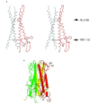
Figure 5. α-Helical interactions and the fusion peptide in HEF2.
a, Stereo diagram of the triple-stranded α-helical bundle of HEF (79–126 of HEF2). Only the region 98–113 interacts across the trimer axis. The red monomer shows residues 4–126 (labelled N, C). b, Trimeric fusion domain of HEF consisting of segments F1, F2, and F3. N terminus of F1 and C terminus of F3 are indicated.

References

    1. Herrler G, Klenk H-D. Structure and function of the HEF glycoprotein of influenza C virus. Adv. Virus Res. 1991;40:213–234. doi: 10.1016/S0065-3527(08)60280-8. - DOI - PMC - PubMed
    1. Wei Y. Anovel variant of the catalytic triad in the Streptomyces scabies esterase. Nature Struct. Biol. 1995;2:218–223. doi: 10.1038/nsb0395-218. - DOI - PubMed
    1. Ho YS. Brain acetylhydrolase that inactivates platelet-activating factor is a G-protein-like trimer. Nature. 1997;385:89–93. doi: 10.1038/385089a0. - DOI - PubMed
    1. Luytjes W, Bredenbeek PJ, Noten AF, Horzinek MC, Spaan WJ. Sequence of mouse hepatitis virus A59 mRNA 2: indications for RNA recombination between coronaviruses and influenza C virus. Virology. 1988;166:415–422. doi: 10.1016/0042-6822(88)90512-0. - DOI - PMC - PubMed
    1. Rosenthal PB. Crystallographic Studies of the Influenza C Virus Surface Glycoprotein. 1996.

Publication types

MeSH terms

Associated data