. 1998 Nov 16;143(4):887-99.
doi: 10.1083/jcb.143.4.887.

Nuclear import and the evolution of a multifunctional RNA-binding protein

Affiliations

Nuclear import and the evolution of a multifunctional RNA-binding protein

J S Rosenblum et al. J Cell Biol. .

Abstract

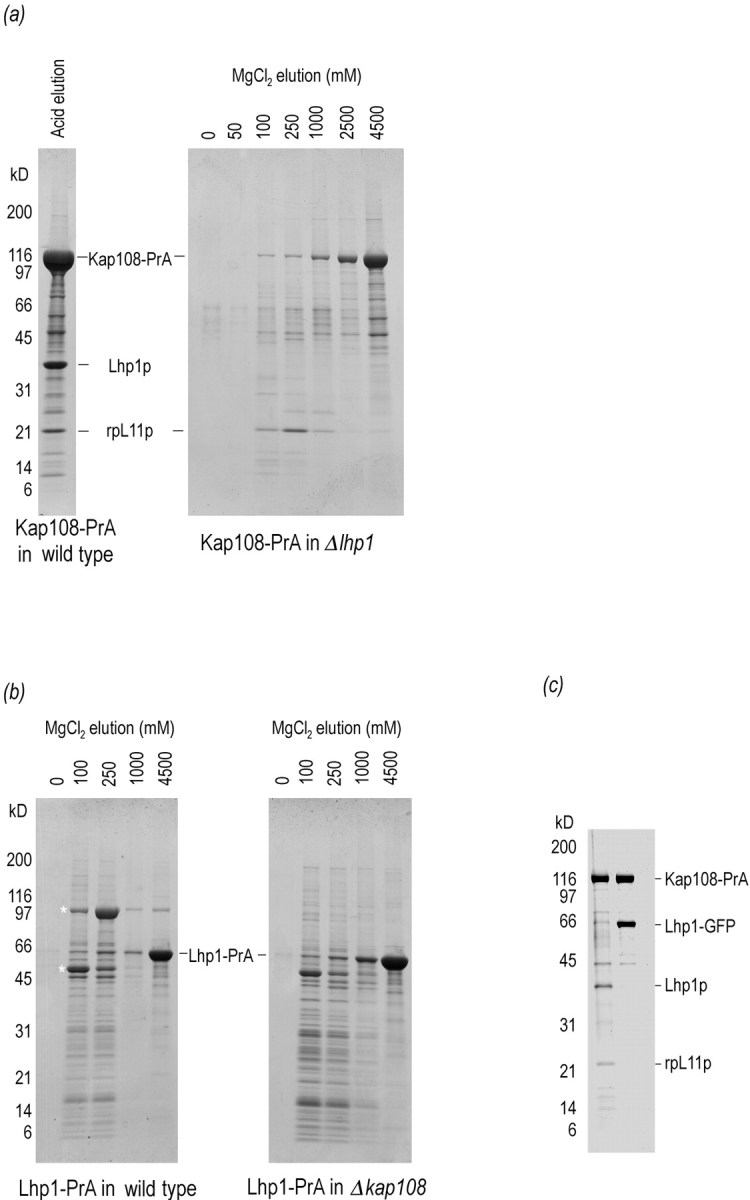
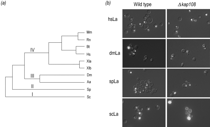
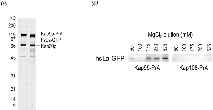
La (SS-B) is a highly expressed protein that is able to bind 3'-oligouridylate and other common RNA sequence/structural motifs. By virtue of these interactions, La is present in a myriad of nuclear and cytoplasmic ribonucleoprotein complexes in vivo where it may function as an RNA-folding protein or RNA chaperone. We have recently characterized the nuclear import pathway of the S. cerevisiae La, Lhp1p. The soluble transport factor, or karyopherin, that mediates the import of Lhp1p is Kap108p/Sxm1p. We have now determined a 113-amino acid domain of Lhp1p that is brought to the nucleus by Kap108p. Unexpectedly, this domain does not coincide with the previously identified nuclear localization signal of human La. Furthermore, when expressed in Saccharomyces cerevisiae, the nuclear localization of Schizosaccharomyces pombe, Drosophila, and human La proteins are independent of Kap108p. We have been able to reconstitute the nuclear import of human La into permeabilized HeLa cells using the recombinant human factors karyopherin alpha2, karyopherin beta1, Ran, and p10. As such, the yeast and human La proteins are imported using different sequence motifs and dissimilar karyopherins. Our results are consistent with an intermingling of the nuclear import and evolution of La.


Figures

Figure 1
Figure 1
Kap108p binds directly to both Lhp1p and rpL11p. The binding sites of Lhp1p and rpL11p on Kap108p overlap. (a) Genomically expressed Kap108– PrA was isolated from cytosol of a wild-type strain (left) and an lhp1-deletion strain (right). After incubation with IgG-Sepharose, unbound proteins were removed and bound proteins were eluted with acid (left) and a MgCl2 gradient (right). After separation on 4–20% SDS-PAGE gels, proteins were visualized with Coomassie blue R. (b) Genomically expressed Lhp1–PrA was isolated from cytosol of a wild-type strain (left) and a KAP108-deletion strain (right). Purified cytosol from each strain was incubated with IgG-Sepharose, washed and eluted with a MgCl2 step gradient. After separation on 4–20% SDS-PAGE gels, proteins were visualized with Coomassie blue R. Asterisk, Lhp1–PrA-associated proteins. (c) Cytosolic Kap108– PrA was isolated from cytosol of a strain without (left lane), and with (right lane), a plasmid expressing an Lhp1– GFP fusion protein. After isolation of Kap108–PrA, bound proteins were eluted with acid. After separation on a 10–20% SDS-PAGE gel, proteins were visualized with Coomassie blue R.
Figure 2
Figure 2
The Kap108p-dependent NLS of Lhp1p overlaps with the RNA recognition motif of Lhp1p. (a) A diagram of the constructs localized by virtue of fusion with GFP. Three relevant features of Lhp1p, the location of the La domain, RRM, and corresponding region of the human La NLS are noted above the bar representing full-length Lhp1p. (b) Approximately equal amounts of total protein from cells expressing the reporter constructs were separated on a 7.5% SDS-PAGE gel and transferred to nitrocellulose. Polyclonal anti-GFP antibodies were used to visualize the fusion proteins. (c) GFP fusion proteins were visualized in live wild-type and Δkap108 cells. Coincident fluorescence and Nomarski optics were used to localize GFP relative to the cell periphery. Fusion proteins were considered nuclear (Nuc) if they were able to concentrate in the nucleus, otherwise they were considered cytoplasmic (Cyt).
Figure 2
Figure 2
The Kap108p-dependent NLS of Lhp1p overlaps with the RNA recognition motif of Lhp1p. (a) A diagram of the constructs localized by virtue of fusion with GFP. Three relevant features of Lhp1p, the location of the La domain, RRM, and corresponding region of the human La NLS are noted above the bar representing full-length Lhp1p. (b) Approximately equal amounts of total protein from cells expressing the reporter constructs were separated on a 7.5% SDS-PAGE gel and transferred to nitrocellulose. Polyclonal anti-GFP antibodies were used to visualize the fusion proteins. (c) GFP fusion proteins were visualized in live wild-type and Δkap108 cells. Coincident fluorescence and Nomarski optics were used to localize GFP relative to the cell periphery. Fusion proteins were considered nuclear (Nuc) if they were able to concentrate in the nucleus, otherwise they were considered cytoplasmic (Cyt).
Figure 3
Figure 3
La proteins localize to the nucleus in S. cerevisiae, but only Lhp1p, the endogenous S. cerevisiae La, needs Kap108p to do so. (a) A phylogenetic tree of the ten cloned La proteins is shown, branches I–IV are indicated. The phylogenetic tree was generated by aligning the NH2-terminal 250 aa of each La protein using ClustalW 1.6 followed by phylogenetic analysis using Phylip (the aligned sequences are available at http://129.85.13.212/LAS.aln). This region contains the La domain and an RRM for each protein. Species abbreviations and references for cDNA sequencing are as follows: Mm, Mus musculus (Topfer et al., 1993); Rn, Rattus norvegicus (Semsei et al., 1993); Bs, Bos taurus (Chan et al., 1989); Hs, Homo sapiens (Chambers et al., 1988); Xla and Xlb, Xenopus laevis (Scherly et al., 1993); Dm, Drosophila melanogaster (Bai et al., 1994); Aa, Aedes albopictus (Pardigon and Strauss, 1996); Sp, Schizosaccharomyces pombe (Van Horn et al., 1997); Sc, Saccharomyces cerevisiae (Yoo and Wolin, 1994). (b) La proteins from the four main branches of the phylogenetic tree in Fig. 3 a were expressed as GFP fusions in S. cerevisiae. Each protein (rows) was expressed in both wild-type (left column) and Δkap108 (right column) cells. Fusion proteins were visualized as described above.
Figure 4
Figure 4
Human La interacts with yeast Kap60p/ Kap95p. (a) Proteins associated with Kap95–PrA were isolated from cytosol of strains without (left) and with (right) a plasmid expressing human La as a GFP fusion (hsLa–GFP). Extracts were passed over an IgG-Sepharose column, which was then washed. Bound proteins were eluted with acid and subjected to SDS-PAGE on a 10–20% gel. Proteins were stained with Coomassie blue R. (b) Proteins associated with Kap95–PrA, (left) and Kap108–PrA, (right), were isolated from cytosol. In both cases, the strains harbored a plasmid expressing hsLa–GFP. In this case, bound proteins were eluted with a MgCl2 gradient, separated by SDS-PAGE, and then transferred to nitrocellulose. The GFP moiety was detected by immunoblotting with anti-GFP antibodies. Only the relevant part of the blot is shown.
Figure 5
Figure 5
Recombinant human Kapα2, Kapβ1, Ran, and p10 reconstitute nuclear import of human La. A bacterially expressed, human La–GFP fusion protein was subjected to the nuclear import assay in permeabilized HeLa cells in the presence of: (first panel) no added factors, (second panel) whole HeLa cytosol, (third panel) Kapα2 and Kapβ1, or (fourth panel) Kapα2, Kapβ1, Ran, and p10.
Figure 6
Figure 6
The extreme COOH termini of La proteins are key elements of their evolution. (a) The final 35 amino acids of ten La proteins (for abbreviations see Fig. 3 legend). White on black background, basic residues; gray on shaded background, acidic residues; gray, other residues. (b) The final 35 amino acids of S. pombe and D. melanogaster were each expressed as fusions with GFP in S. cerevisiae. The fusion constructs were visualized as previously described.
Figure 7
Figure 7
A representation of important domains for four evolutionarily distinct La proteins. Bars proportional to the length of the human, Drosophila, S. pombe, and S. cerevisiae La proteins are shown. Black lines, RRMs; two-headed arrows, proteins NLSs. The ATP-binding domain of human La is indicated.

References

    1. Aitchison JD, Rout MP, Marelli M, Blobel G, Wozniak RW. Two novel related yeast nucleoporins Nup170p and Nup157p: complementation with the vertebrate homologue Nup155p and functional interactions with the yeast nuclear pore-membrane protein Pom152p. J Cell Biol. 1995;131:1133–1148. - PMC - PubMed
    1. Aitchison JD, Blobel G, Rout MP. Kap104p: a karyopherin involved in the nuclear transport of messenger RNA binding proteins. Science. 1996;274:624–627. - PubMed
    1. Albertini, M., L.F. Pemberton, J.S. Rosenblum, and G. Blobel. 1998. Nuclear import and the evolution of a multifunctional RNA-binding protein. J. Cell Biol. In press. - PMC - PubMed
    1. Ali N, Siddiqui A. The La antigen binds 5′ noncoding region of the hepatitis C virus RNA in the context of the initiator AUG codon and stimulates internal ribosome entry site-mediated translation. Proc Natl Acad Sci USA. 1997;94:2249–2254. - PMC - PubMed
    1. Ausubel, F.M., R. Brent, R.E. Kingston, D.D. Moore, J.A. Smith, J.G. Seidman, and K. Struhl. 1997. Current Protocols in Molecular Biology. Vol. 2. John Wiley & Sons, Inc., New York. 13.0.1–13.13.9.

Publication types

MeSH terms