. 1998 Dec;36(12):3552-7.
doi: 10.1128/JCM.36.12.3552-3557.1998.

Genetic variability among group A and group B respiratory syncytial viruses in a children's hospital

Affiliations

Genetic variability among group A and group B respiratory syncytial viruses in a children's hospital

W B Coggins et al. J Clin Microbiol. 1998 Dec.

Abstract

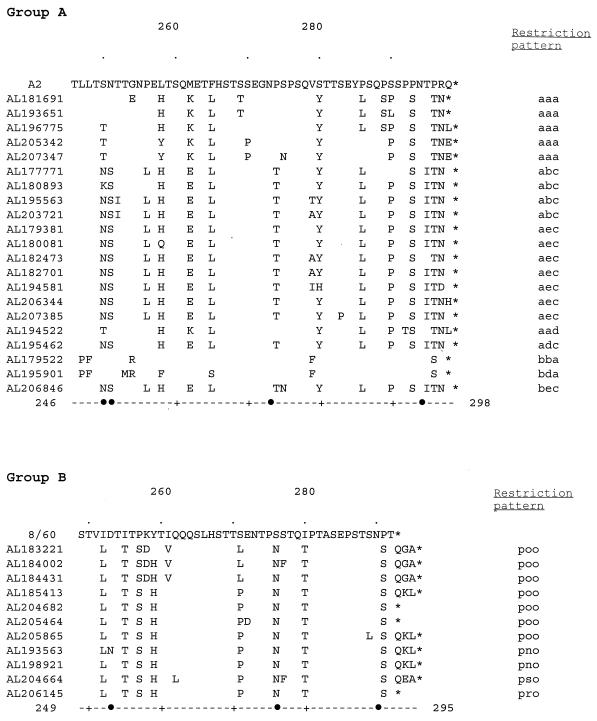
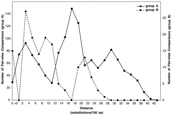
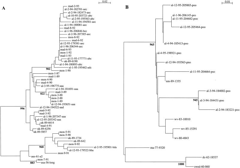
Respiratory syncytial (RS) viruses isolated over three epidemic periods in a children's hospital in the United States were analyzed. The viruses (n = 174) were characterized as to major antigenic group (group A or B) by a PCR-based assay. Group A RS viruses were dominant the first 2 years, followed by a year with group B dominance (ratios of group A to group B viruses for epidemic periods, 56/4 for 1993-1994, 42/3 for 1994-1995, and 19/50 for 1995-1996). Genetic variability within the groups was assessed by restriction fragment analysis of PCR products; 79 isolates were also analyzed by nucleotide sequence determination of a variable region of the glycoprotein G gene. Among the group A RS virus isolates, this G-protein variable region had amino acid differences of as great as 38%. The G-protein amino acids of the group A viruses differed by up to 31% from the G-protein amino acids of a prototype (A2) group A virus. Among the group B RS virus G proteins, amino acid differences were as great as 14%. The G-protein amino acids of the group B viruses differed by up to 27% from the G-protein amino acids of a prototype (18537) group B virus. The group A and group B RS viruses demonstrated genetic variability between years and within individual years. Phylogenetic analysis revealed that there were multiple evolutionary lineages among both the group A and group B viruses. Among the recent group B isolates, variability was less than that seen for the group A viruses. However, comparisons to prototype strains revealed that the group B RS viruses may vary more extensively than was observed over the 3 years studied in the present investigation.


Figures

FIG. 1
FIG. 1
Alignment of the deduced amino acid sequences of G proteins of the group A (A) and group B (B) isolates from a children’s hospital. The sequences from amino acids 246 (group A) or 249 (group B) to the end of the G protein are shown in comparison with the sequence of prototype isolate A2 (A) or 8/60 (B). Differences in the sequences of the isolates from clinical samples in comparison to the sequences of the prototype isolates are shown. The name of each virus isolated in the Children’s Hospital of Alabama begins each line, and restriction patterns are shown at the end of the sequence. Symbols: •, potential N-linked glycosylation sites in any of the sequences; ∗, termination codons. The residues are indicated by numbers at the ends of the dashed lines at the bottom; dots above the sequences indicate 10-residue increments, with residues 260 and 280 being shown.
FIG. 2
FIG. 2
Pairwise distances among group A (A) and group B (B) RS virus G proteins. All possible pairwise comparisons were made among amino acid sequences from isolates at the children’s hospital and published human RS virus G-protein sequences available through GenBank for the region of the G protein analyzed in this report. See Fig. 3 for a list of the published sequences which were used here. The distances represent the number of substitutions/100 amino acids (aa) for each pairwise comparison. No correction was used for multiple substitutions at single sites. The figure plots the number of pairwise sequence comparisons that have distance measurements within each indicated range. Sequence distances for group A and group B viruses are shown separately by using different scales for the number of sequence comparisons.
FIG. 3
FIG. 3
Group A (A) and B (B) RS virus G-protein phylogenetic relationships. Partial G-protein sequences from RS viruses isolated at the Children’s Hospital of Alabama were compared to published G-protein sequences available through GenBank. Viruses are identified by the geographic location (from the United States, al, Alabama; wv, West Virginia [23]; dc, District of Columbia [16], ma, Massachusetts [23]; md, Maryland [16], and nm, New Mexico [23]; from the United Kingdom, uk [5]; from Spain, mad, Madrid; from Uruguay, mon, Montevideo [12]; from Sweden, swed [22]; and from Australia, aus [28]), year or month and year of isolation, and, for isolates from the United States and the United Kingdom a number designation. For isolates from Alabama a three-letter designation that describes the restriction pattern observed after restriction endonuclease digestion of the PCR DNA products follows the designations described above. A single sequence was used for each unique nucleotide sequence within each restriction fragment pattern among the isolates described here (Table 1). The nucleotide sequence alignments of either group A or group B sequences were used to create the neighbor-joining trees displayed in the figure. The scales represent either 0.02 (group A) or 0.01 (group B) substitutions per base per indicated horizontal distance. The numbers present at some of the internal nodes of the trees represent the number of bootstrap replicates of 1,000 that display the indicated sequence groupings. Only significant bootstrap replicate numbers with values of greater than 800 are shown.

References

    1. Air G M, Gibbs A J, Laver W, Webster R G. Evolutionary changes in influenza B are not primarily governed by antibody selection. Proc Natl Acad Sci USA. 1990;87:3884–3888. - PMC - PubMed
    1. Anderson L J, Hendry R M, Pierik L T, Tsou C, McIntosh K. Multicenter study of strains of respiratory syncytial virus. J Infect Dis. 1991;163:687–692. - PubMed
    1. Anderson L J, Hierholzer J C, Tsou C, Hendry R M, Fernie B F, Stone Y, McIntosh K. Antigenic characterization of respiratory syncytial virus strains with monoclonal antibodies. J Infect Dis. 1985;151:626–633. - PubMed
    1. Cane P A. Analysis of linear epitopes recognised by the primary human antibody response to a variable region of the attachment (G) protein of respiratory syncytial virus. J Med Virol. 1997;51:297–304. - PubMed
    1. Cane P A, Matthews D A, Pringle C R. Identification of variable domains of the attachment (G) protein of subgroup A respiratory syncytial viruses. J Gen Virol. 1991;72:2091–2096. - PubMed

Publication types

LinkOut - more resources