. 1998 Dec;18(12):6921-9.
doi: 10.1128/MCB.18.12.6921.

Protein kinase A-dependent derepression of the human prodynorphin gene via differential binding to an intragenic silencer element

Affiliations

Protein kinase A-dependent derepression of the human prodynorphin gene via differential binding to an intragenic silencer element

A M Carrión et al. Mol Cell Biol. 1998 Dec.

Abstract

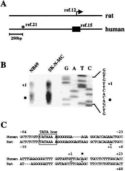
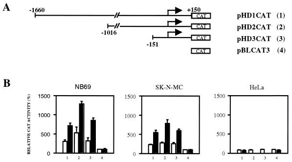
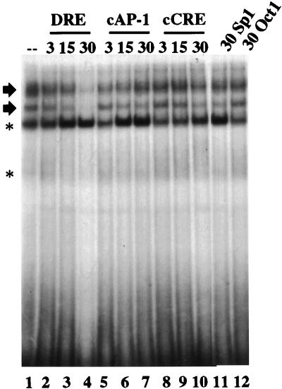
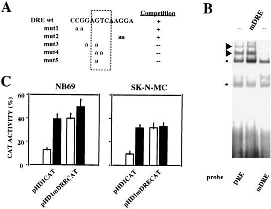
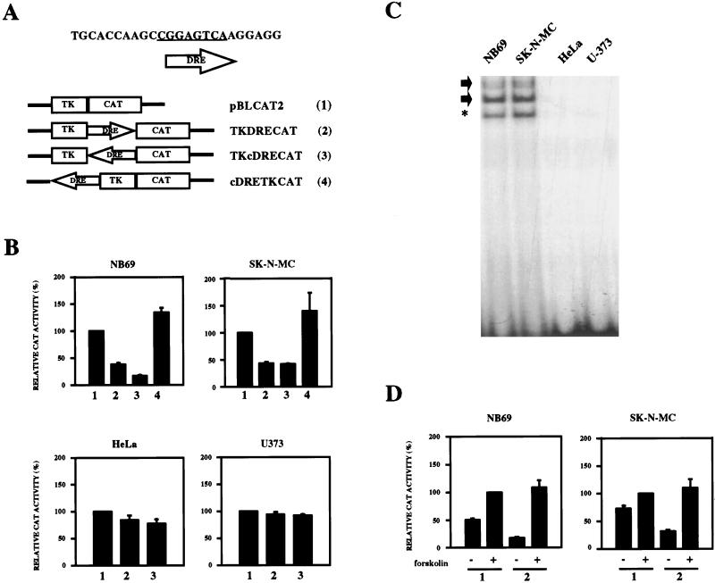
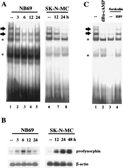
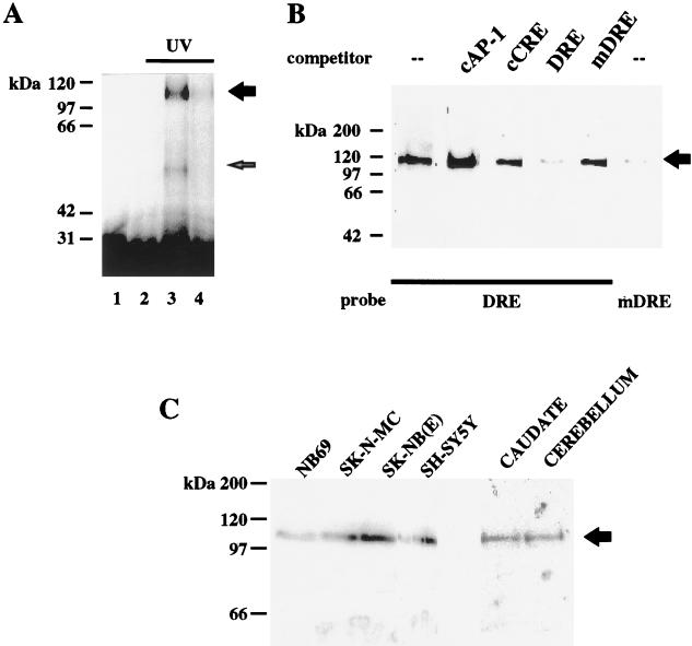
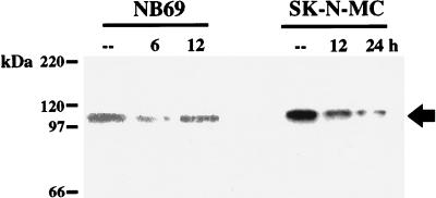
Induction of the prodynorphin gene has been implicated in medium and long-term adaptation during memory acquisition and pain. By 5' deletion mapping and site-directed mutagenesis of the human prodynorphin promoter, we demonstrate that both basal transcription and protein kinase A (PKA)-induced transcription in NB69 and SK-N-MC human neuroblastoma cells are regulated by the GAGTCAAGG sequence centered at position +40 in the 5' untranslated region of the gene (named the DRE, for downstream regulatory element). The DRE repressed basal transcription in an orientation-independent and cell-specific manner when placed downstream from the heterologous thymidine kinase promoter. Southwestern blotting and UV cross-linking experiments with nuclear extracts from human neuroblastoma cells or human brain revealed a protein complex of approximately 110 kDa that specifically bound to the DRE. Forskolin treatment reduced binding to the DRE, and the time course paralleled that for an increase in prodynorphin gene expression. Our results suggest that under basal conditions, expression of the prodynorphin gene is repressed by occupancy of the DRE site. Upon PKA stimulation, binding to the DRE is reduced and transcription increases. We propose a model for human prodynorphin activation through PKA-dependent derepression at the DRE site.


Figures

FIG. 1
FIG. 1
The transcription start site in the human prodynorphin gene. (A) Alignment of the rat and human prodynorphin genes. The arrow indicates the transcription start site in the rat gene (12). The box encompasses the region in the human gene recently proposed to contain the start of transcription for the human gene (15), and the asterisk denotes a position proposed earlier (21). (B) Primer extension analysis of human prodynorphin transcripts using mRNA from NB69 and SK-N-MC cells. The first transcription start site is represented by +1, and the solid circle indicates a second transcription start site 5 bp downstream. A sequencing reaction with the primer used for the extension analysis is shown to the right. (C) Nucleotide sequence of the region in the human prodynorphin gene containing the transcription start site (+1 and solid circle) shown in relation to a putative TATA box (boxed) located at −50 bp. For comparison, the corresponding region of the rat gene is shown.
FIG. 2
FIG. 2
Deletion mapping of the human prodynorphin promoter. (A) Schematic representation of the 5′ deletions (no. 1 to 3) in the human promotor and the reporter vector pBLCAT3 (no. 4). (B) Transient transfections in the human neuroblastoma cells NB69 and SK-N-MC and in HeLa cells with the reporter plasmids 1 to 4 shown in panel A, corresponding to the numbers on the x axis. The results are expressed as CAT activity relative to that of empty reporter vector pBLCAT3 (no. 4) under basal conditions. Open bars represent values from untreated cultures, and solid bars represent values from forskolin-treated cultures. The transfection experiments were repeated seven times in duplicate.
FIG. 3
FIG. 3
Electrophoretic mobility shift analysis of the DRE from the human prodynorphin gene. Results from competition assays of the DRE retarded bands with the indicated fold excess (3-, 15-, and 30-fold) of cold competitors (top) are shown. The two specific DRE retarded bands are indicated by arrows. Nonspecific retarded bands are indicated by asterisks.
FIG. 4
FIG. 4
Site-directed mutagenesis of the DRE on the human prodynorphin promoter. (A) Sequence of the different DRE mutant oligonucleotides aligned with the wild-type DRE. Their ability to compete the specific DRE retarded bands is shown to the right. (B) Electrophoretic mobility shift assay showing the retardation pattern with the wild-type DRE as a probe (lane 1) and the effect on the specific retarded bands (arrowheads) of the selected mutation DREmut5 (renamed mDRE [lane 3]). Competition with 30-fold excess of cold mutated DRE (mDRE) is shown in lane 2. Asterisks denote nonspecific retarded bands. (B) Transient transfections in NB69 and in SK-N-MC cells were performed with plasmids containing either the wild-type promoter (pHD1CAT) or the promoter mutated at the DRE site (pHD1mDRECAT). CAT activity obtained from each reporter plasmid under basal conditions (open bars) and after forskolin treatment (solid bars) is shown. The transfection experiments were repeated four times in duplicate.
FIG. 5
FIG. 5
Repression of the heterologous promoter from the thymidine kinase gene by DRE. (A) Schematic representation of the DRE constructs used (no. 2 to 4) and the empty reporter vector pBLCAT2 (no. 1). (B) Effect of the DRE under basal conditions after transient transfections in NB69, SK-N-MC, HeLa, and U373 cell lines with the reporter plasmids 2 to 4 shown in panel A, corresponding to the numbers on the x axis. The results are expressed as CAT activity relative to that of the pBLCAT2 reporter (no. 1). (C) Electrophoretic mobility shift assay with the DRE as a probe and nuclear extracts from the indicated cell lines (top). Arrows indicate the DRE-specific retarded bands. Asterisks denote nonspecific retarded bands. (D) Effect of the DRE after transient transfections in forskolin-treated NB69 and SK-N-MC cells using the reporter plasmids 1 and 2 shown in panel A, corresponding to the numbers on the x axis. The results are expressed as CAT activity relative to that of the pBLCAT2 reporter after forskolin treatment. All transfection experiments were repeated four times in duplicate.
FIG. 6
FIG. 6
Blocking of binding to the DRE after PKA activation correlates with the increase in prodynorphin expression in NB69 and SK-N-MC cells. (A) Electrophoretic mobility shift assays showing reduction in the DRE-specific retarded bands (arrows) at the indicated times (top) after forskolin treatment. (B) Southern blot after RT-PCR showing increase in prodynorphin expression in NB69 and SK-N-MC cells at the indicated times (top) after forskolin treatment. Amplification of β-actin was performed in parallel as a control for the mRNA input. (C) Electrophoretic mobility shift assay showing reduction in the DRE-specific retarded bands (arrows) by dibutyryl-cAMP (lane 2) or forskolin (lane 3) treatments at the 6-h time point with the NB69 cell line. The effect of forskolin treatment blocked by H89 is shown in lane 4. Asterisks in panels A and C denote nonspecific retarded bands.
FIG. 7
FIG. 7
Characterization of the nuclear activity that binds to the DRE site. (A) NB69 nuclear extracts were UV cross-linked after incubation with the DRE probe and electrophoresed. Three micrograms of proteinase K (lane 2) or a 30-fold excess of cold DRE (lane 4) abolished or reduced, respectively, the specific autoradiographic signal (lane 3). Without UV irradiation, no specific band was observed (lane 1). (B) Southwestern analysis with NB69 nuclear extracts. A protein complex of 110 kDa was obtained with the DRE probe. The autoradiographic band was competed with an excess of cold DRE but not by cold mDRE (top). A probe containing the mutated DRE seriously reduced the autoradiographic signal. (C) Southwestern analysis with the DRE as a probe and nuclear extracts from the neuroblastoma cell lines indicated (top) and human brain samples. The arrow indicates the specific band obtained for the 110-kDa protein complex. Protein molecular mass markers are shown to the left.
FIG. 8
FIG. 8
PKA activation blocks binding of the DRE probe to the 110-kDa protein complex. The results represent Southwestern analysis with nuclear extracts from NB69 and SK-N-MC cells at different times after forskolin treatment. The arrow indicates the specific band obtained for the 110-kDa protein complex.

References

    1. Adams A D, Choate D M, Thompson M A. NF1-L is the DNA-binding component of the protein complex at the peripherin negative regulatory element. J Biol Chem. 1995;270:6975–6983. - PubMed
    1. Alvarez G, Fairén A, Douglass J, Naranjo J R. Expression of the prodynorphin gene in the developing and adult cerebral cortex of the rat: an in situ hybridization study. J Comp Neurol. 1990;300:287–300. - PubMed
    1. Andrews N C, Faller D V. A rapid micropreparation technique for extraction of DNA-binding proteins from limiting numbers of mammalian cells. Nucleic Acids Res. 1991;19:2499. - PMC - PubMed
    1. Ausubel F M, Brent R, Kingston R E, Moore D D, Seidman J G, Smith J A, Struhl K. Current protocols in molecular biology. New York, N.Y: Wiley; 1995.
    1. Bessis A, Champtiaux N, Chatelin L, Changeux J-P. The neuron-restrictive silencer element: a dual enhancer/silencer crucial for patterned expression of a nicotine receptor gene in the brain. Proc Natl Acad Sci USA. 1997;94:5906–5911. - PMC - PubMed

Publication types

MeSH terms

Associated data

LinkOut - more resources