. 1998 Dec;18(12):6951-61.
doi: 10.1128/MCB.18.12.6951.

Nucleophosmin-anaplastic lymphoma kinase of large-cell anaplastic lymphoma is a constitutively active tyrosine kinase that utilizes phospholipase C-gamma to mediate its mitogenicity

Affiliations

Nucleophosmin-anaplastic lymphoma kinase of large-cell anaplastic lymphoma is a constitutively active tyrosine kinase that utilizes phospholipase C-gamma to mediate its mitogenicity

R Y Bai et al. Mol Cell Biol. 1998 Dec.

Abstract

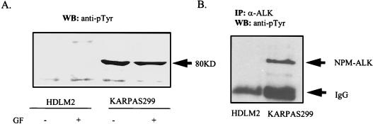
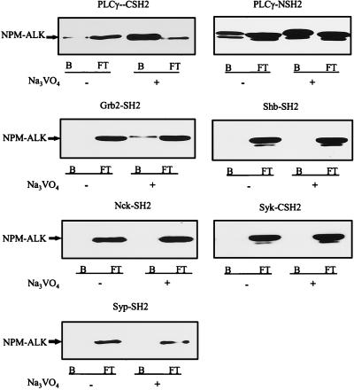
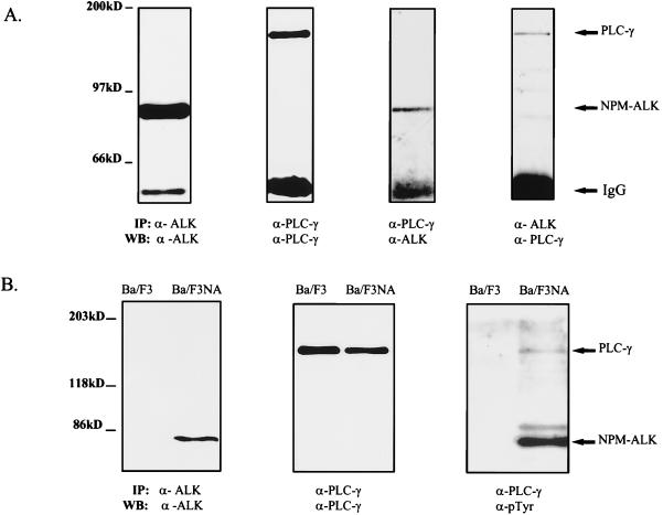
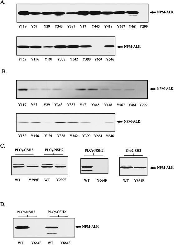
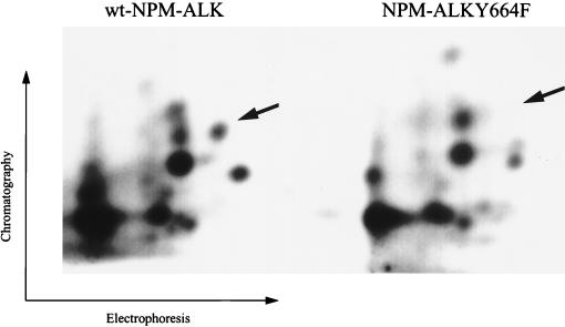
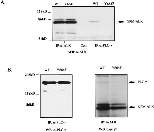
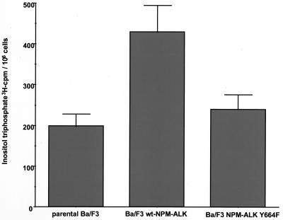
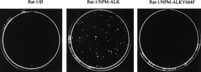
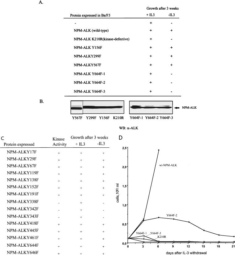
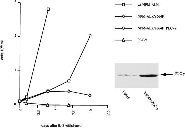
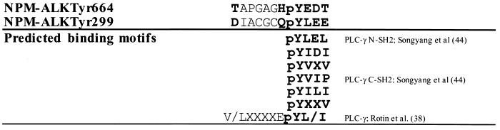
Large-cell anaplastic lymphoma is a subtype of non-Hodgkin's lymphoma characterized by the expression of CD30. More than half of these lymphomas have a chromosomal translocation, t(2;5), that leads to the expression of a hybrid protein comprised of the nucleolar phosphoprotein nucleophosmin (NPM) and the anaplastic lymphoma kinase (ALK). Here we show that transfection of the constitutively active tyrosine kinase NPM-ALK into Ba/F3 and Rat-1 cells leads to a transformed phenotype. Oncogenic tyrosine kinases transform cells by activating the mitogenic signal transduction pathways, e.g., by binding and activating SH2-containing signaling molecules. We found that NPM-ALK binds most specifically to the SH2 domains of phospholipase C-gamma (PLC-gamma) in vitro. Furthermore, we showed complex formation of NPM-ALK and PLC-gamma in vivo by coimmunoprecipitation experiments in large-cell anaplastic lymphoma cells. This complex formation leads to the tyrosine phosphorylation and activation of PLC-gamma, which can be corroborated by enhanced production of inositol phosphates (IPs) in NPM-ALK-expressing cells. By phosphopeptide competition experiments, we were able to identify the tyrosine residue on NPM-ALK responsible for interaction with PLC-gamma as Y664. Using site-directed mutagenesis, we constructed a comprehensive panel of tyrosine-to-phenylalanine NPM-ALK mutants, including NPM-ALK(Y664F). NPM-ALK(Y664F), when transfected into Ba/F3 cells, no longer forms complexes with PLC-gamma or leads to PLC-gamma phosphorylation and activation, as confirmed by low IP levels in these cells. Most interestingly, Ba/F3 and Rat-1 cells expressing NPM-ALK(Y664F) also show a biological phenotype in that they are not stably transformed. Overexpression of PLC-gamma can partially rescue the proliferative response of Ba/F3 cells to the NPM-ALK(Y664F) mutant. Thus, PLC-gamma is an important downstream target of NPM-ALK that contributes to its mitogenic activity and is likely to be important in the molecular pathogenesis of large-cell anaplastic lymphomas.


Figures

FIG. 1
FIG. 1
Growth factor-independent autophosphorylation of NPM-ALK in large-cell anaplastic lymphoma cells. (A) HDLM2 and Karpas299 cells were cultured with 0.5% (−) or 10% (+) FCS for 24 h. Lysates from 107 cells were prepared as described in Materials and Methods and subjected to SDS-PAGE, and immunoblotting was performed with the antiphosphotyrosine antibody PY20. (B) HDLM2 and Karpas299 cells (107 each) were lysed and immunoprecipitated with anti-ALK antibody. Immunoblotting was performed with the antiphosphotyrosine antibody 4G10. GF, growth factor; IP, immunoprecipitation; WB, Western blotting; IgG, immunoglobulin G.
FIG. 2
FIG. 2
The SH2 domains of PLC-γ and Grb2 bind to NPM-ALK from large-cell anaplastic lymphoma cells. For each binding assay, 107 Karpas299 cells were cultured without (−) or with (+) 100 μM Na3VO4 overnight. Cells were washed briefly with ice-cold PBS, lysed in 0.5% Triton X-100-containing lysis buffer, and incubated with different GST-SH2 fusion proteins for 1 h at 4°C. The bound (B) and the flowthrough (FT) fractions were collected with glutathione-Sepharose, subjected to SDS–7.5% PAGE, and analyzed by anti-ALK immunoblotting.
FIG. 3
FIG. 3
NPM-ALK association with PLC-γ in vivo leads to tyrosine phosphorylation of PLC-γ in NPM-ALK-expressing cells. (A) Karpas299 cells (5 × 10⁶ [two left panels] or 2 × 10⁷ [two right panels]) were subjected to immunoprecipitations and immunoblotting with the antibodies indicated. Association of NPM-ALK with PLC-γ was demonstrated by coprecipitation of NPM-ALK with an anti-PLC-γ antibody (second panel from right) and by coprecipitation of PLC-γ with anti-ALK antibody (right panel). (B) The lymphocyte cell line Ba/F3 was transformed with wt NPM-ALK in pCDNA3 by electroporation, and stable clones were established by selection in G418 for 2 weeks, as described in Materials and Methods. Parental Ba/F3 cells and Ba/F3 cells expressing NPM-ALK (Ba/F3NA) (107 each) were analyzed by immunoprecipitation and immunoblotting with anti-ALK antibody to demonstrate the expression of wt NPM-ALK in Ba/F3NA cells (left panel) or with anti-PLC-γ antibody to examine endogenous PLC-γ expression (middle panel). Ba/F3 and Ba/F3NA cells (4 × 10⁷ each) were lysed and immunoprecipitated with anti-PLC-γ antibody, followed by immunoblotting with antiphosphotyrosine antibody (PY20 and 4G10) (right panel). Tyrosine phosphorylation and coprecipitation of PLC-γ with NPM-ALK were detected only in NPM-ALK-expressing cells (Ba/F3NA). IP, immunoprecipitation; WB, Western blotting; IgG, immunoglobulin G.
FIG. 4
FIG. 4
Phosphopeptide competition identifies Tyr664 in NPM-ALK as the binding site for PLC-γ. (A and B) Tyrosine-phosphorylated peptides corresponding to the putative autophosphorylation sites in NPM-ALK were synthesized with an SMPS 350 (Zinsser Analytik, Frankfurt, Germany) according to the method of Atherton and Sheppard (2). Tyrosine-phosphorylated peptides (200 μM) were incubated with ∼3 μg of GST fusion proteins of the PLC-γ N-terminal (A) or C-terminal (B) SH2 domain in lysis buffer for 1 h at 4°C. Cell lysates of 106 Karpas299 cells were then added to each binding reaction mixture, and mixtures were incubated for a further hour. Complexes were finally precipitated with glutathione-Sepharose, and samples were subjected to SDS–7.5% PAGE and analyzed by anti-ALK immunoblotting. Peptides Y299 and Y664 completely blocked in vitro association of NPM-ALK and the N-terminal and C-terminal SH2 domains of PLC-γ. (C) wt NPM-ALK (WT) and NPM-ALK mutants Y299F and Y664F were translated in vitro and labeled with [35S]methionine by the TNT coupled reticulocyte lysate system with T7 RNA polymerase. In vitro binding with the GST-SH2 domains of PLC-γ and Grb2 was performed as described in Material and Methods with ∼3 μg of GST fusion protein. Samples were resolved by SDS–7.5% PAGE and visualized by autoradiography. Y664, but not Y299, is essential for the binding of PLC-γ to NPM-ALK. (D) wt NPM-ALK (WT) and NPM-ALK(Y664F) sequences in pCDNA3 were stably transfected into Ba/F3 cells. Cell lysates were incubated with PLC-γ GST-NSH2 and PLC-γ GST-CSH2 and precipitated with glutathione-Sepharose, and samples were subjected to SDS–7.5% PAGE and analysis by anti-ALK immunoblotting.
FIG. 5
FIG. 5
Two-dimensional tryptic phosphopeptide maps of wt NPM-ALK and NPM-ALK(Y664F). In vivo-labeled wt NPM-ALK- and NPM-ALK(Y664F)-expressing Ba/F3 cells were prepared as described in Materials and Methods. NPM-ALK proteins were immunoprecipitated and separated by SDS-PAGE. The gel fragments containing NPM-ALK were then extensively digested with trypsin. The resulting digests were washed three times with water and then separated on a TLC cellulose plate electrophoretically at pH 1.9, followed by ascending chromatography (1-butanol–pyridine–acetic acid–water, 375:250:75:300). Radiolabeled phosphopeptides were visualized by autoradiography. The phosphopeptides of wt NPM-ALK and NPM-ALK(Y664F) were loaded on the same TLC plate and separated by a distance of 10 cm. Arrows indicate the position of the phosphopeptide spot missing in the map of the NPM-ALK(Y664F) mutant.
FIG. 6
FIG. 6
In vivo association and tyrosine phosphorylation of PLC-γ by NPM-ALK require Tyr664. (A) Coprecipitation of PLC-γ with NPM-ALK was examined in lysates of 5 × 10⁷ Ba/F3 cells stably transfected with wt NPM-ALK (WT) and NPM-ALK(Y664F). Immunoprecipitation was performed with anti-ALK antibody, rabbit anti-mouse antibody (Con.), or anti-PLC-γ antibody. Samples were resolved by SDS–7.5% PAGE and analyzed by immunoblotting with anti-ALK antibody. (B) Expression levels of PLC-γ (left panel) and tyrosine phosphorylation of NPM-ALK and PLC-γ (right panel) in Ba/F3 cells transfected with wt NPM-ALK and NPM-ALK(Y664F) were determined by immunoprecipitation and immunoblotting with the antibodies indicated. IP, immunoprecipitation; WB, Western blotting.
FIG. 7
FIG. 7
IP levels in Ba/F3 cells transfected with wt NPM-ALK and mutant Y664F. Ba/F3 cells (5 × 10⁶) stably expressing wt NPM-ALK or NPM-ALK(Y664F) were incubated for 24 h in RPMI medium containing 10% FCS and 2 μCi of myo-[3H]inositol. The media were then removed, and the cells were washed thoroughly and incubated in 0.5 ml of PBS solution containing 10 mM LiCl. After 1 h the incubation was stopped by adding 250 μl of 3.5% HCl, and the cell lysates were frozen at −80°C. The IPs formed were determined as described in Materials and Methods. Data represent means ± standard deviations from three independent experiments.
FIG. 8
FIG. 8
Rat-1 soft-agar assay of wt NPM-ALK and NPM-ALK(Y664F). The transfections of Rat-1 cells with pCDNA3 empty vector, wt NPM-ALK, and NPM-ALK(Y664F) mutant were performed with the DOTAP transfection reagent. After undergoing selection with G418 (0.75 mg/ml), the cells were used for Rat-1 soft-agar assays. On each 6-cm dish 2 × 10⁴ cells were plated, and representative soft-agar plates were photographed after 14 days.
FIG. 9
FIG. 9
The transforming potential of wt NPM-ALK and Tyr-to-Phe NPM-ALK mutants in Ba/F3 cells. (A) Ba/F3 cells were stably transfected with the pCDNA3 empty vector (−) or with vector expressing wt or mutant NPM-ALK by electroporation and selection in RPMI medium containing 1 mg of G418 per ml, 10% FCS, and 1.5 ng of murine IL-3 per ml. Three single-cell-derived clones of Ba/F3 cells transfected with NPM-ALK(Y664F) (Y664F 1, 2, and 3) were obtained by limiting dilution. Growth with or without IL-3 was monitored over a period of 3 weeks, with viable cells determined by using trypan blue staining and a hemacytometer. (B) Expression levels of the different NPM-ALK constructs were determined by SDS–7.5% PAGE and anti-ALK immunoblotting. WB, Western blotting. (C) Tyr-to-Phe mutants were stably expressed in Ba/F3 cells. Protein expression and kinase activity of the mutants were verified by immunoblotting (data not shown). Growth with or without IL-3 was monitored over a period of 3 weeks, with viable cells determined by using trypan blue staining and a hemacytometer. (D) After IL-3 withdrawal, cell numbers in each culture were determined by using trypan blue staining and a hemacytometer. Cell culture media were renewed every 6 days.
FIG. 10
FIG. 10
Overexpression of a PLC-γ cDNA rescues Ba/F3 NPM-ALK(Y664F) cells for IL-3-independent growth. The PLC-γ2 sequence in pCDNA3.1 Zeo(−) was transfected into parental Ba/F3 and NPM-ALK(Y664F) 2-expressing Ba/F3 cells, followed by selection in RPMI medium containing 0.25 mg of zeocin per ml, 10% FCS, and 1.5 ng of murine IL-3 per ml. Growth without IL-3 was monitored by determining viable cells by using trypan blue staining and a hemacytometer. The experiment was performed four times with two independent PLC-γ transfections, and results of a representative experiment are shown. The expression level of PLC-γ in the cell lysates was determined by SDS-PAGE and immunoblotting with anti-PLC-γ2 antibody.
FIG. 11
FIG. 11
Amino acid sequences surrounding Tyr664 and Tyr299 of NPM-ALK and the predicted binding motifs for PLC-γ SH2 domains.

References

    1. Alimandi M, Heidaran M A, Gutkind J S, Zhang J, Ellmore N, Valius M, Kazlauskas A, Pierce J H, Li W. PLC-gamma activation is required for PDGF-betaR-mediated mitogenesis and monocytic differentiation of myeloid progenitor cells. Oncogene. 1997;15:585–593. - PubMed
    1. Atherton E, Sheppard R C. Solid phase peptide synthesis—a practical approach. Oxford, England: IRL Press; 1989.
    1. Bartram C R, de Klein A, Hagemeijer A, van Agthoven T, van Kessel A D, Bootsma D, Grosveld G, Ferguson-Smith M A, Davies T, Stone M, Heisterkamp N, Stephenson J R, Groffen J. Translocation of c-Abl oncogene correlates with the presence of a Philadelphia chromosome in chronic myelocytic leukemia. Nature. 1983;306:277–280. - PubMed
    1. Bischof D, Pulford K, Mason D Y, Morris S W. Role of the nucleophosmin (NPM) portion of the non-Hodgkin’s lymphoma-associated NPM-anaplastic lymphoma kinase fusion protein in oncogenesis. Mol Cell Biol. 1997;17:2312–2325. - PMC - PubMed
    1. Borer R A, Lehner C F, Eppenberger H M, Nigg E A. Major nucleolar proteins shuttle between nucleus and cytoplasm. Cell. 1989;56:379–390. - PubMed

Publication types

MeSH terms