. 1998 Dec;18(12):6995-7008.
doi: 10.1128/MCB.18.12.6995.

Regulation of RasGRP via a phorbol ester-responsive C1 domain

Affiliations

Regulation of RasGRP via a phorbol ester-responsive C1 domain

C E Tognon et al. Mol Cell Biol. 1998 Dec.

Abstract

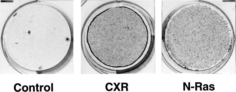
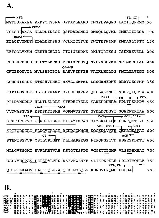
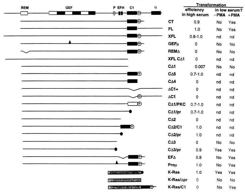
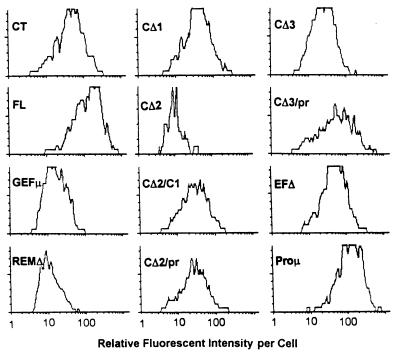
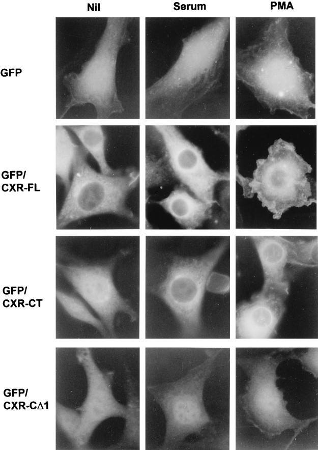
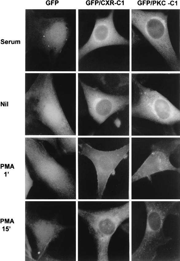
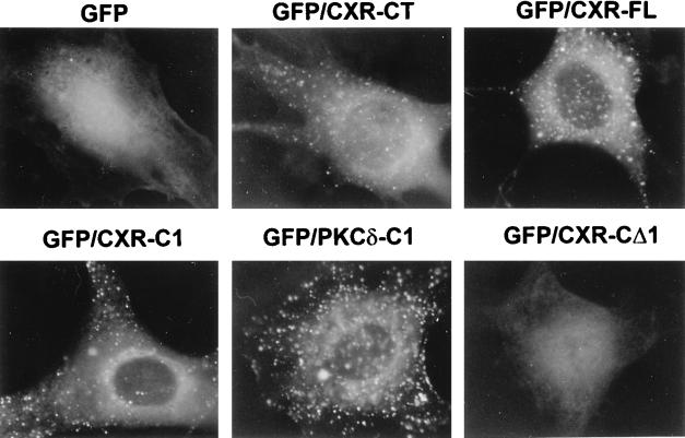
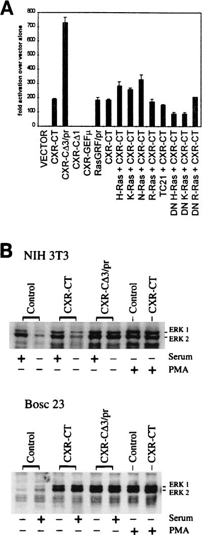
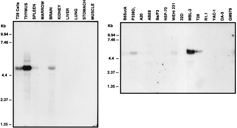
As part of a cDNA library screen for clones that induce transformation of NIH 3T3 fibroblasts, we have isolated a cDNA encoding the murine homolog of the guanine nucleotide exchange factor RasGRP. A point mutation predicted to prevent interaction with Ras abolished the ability of murine RasGRP (mRasGRP) to transform fibroblasts and to activate mitogen-activated protein kinases (MAP kinases). MAP kinase activation via mRasGRP was enhanced by coexpression of H-, K-, and N-Ras and was partially suppressed by coexpression of dominant negative forms of H- and K-Ras. The C terminus of mRasGRP contains a pair of EF hands and a C1 domain which is very similar to the phorbol ester- and diacylglycerol-binding C1 domains of protein kinase Cs. The EF hands could be deleted without affecting the ability of mRasGRP to transform NIH 3T3 cells. In contrast, deletion of the C1 domain or an adjacent cluster of basic amino acids eliminated the transforming activity of mRasGRP. Transformation and MAP kinase activation via mRasGRP were restored if the deleted C1 domain was replaced either by a membrane-localizing prenylation signal or by a diacylglycerol- and phorbol ester-binding C1 domain of protein kinase C. The transforming activity of mRasGRP could be regulated by phorbol ester when serum concentrations were low, and this effect of phorbol ester was dependent on the C1 domain of mRasGRP. The C1 domain could also confer phorbol myristate acetate-regulated transforming activity on a prenylation-defective mutant of K-Ras. The C1 domain mediated the translocation of mRasGRP to cell membranes in response to either phorbol ester or serum stimulation. These results suggest that the primary mechanism of activation of mRasGRP in fibroblasts is through its recruitment to diacylglycerol-enriched membranes. mRasGRP is expressed in lymphoid tissues and the brain, as well as in some lymphoid cell lines. In these cells, RasGRP has the potential to serve as a direct link between receptors which stimulate diacylglycerol-generating phospholipase Cs and the activation of Ras.


Figures

FIG. 1
FIG. 1
Transforming activity of the CXR-CT cDNA clone encoding truncated mRasGRP. NIH 3T3 cells were plated at low density and infected with CTV83B, which is an empty retroviral vector (Control); with retroviral vector CTV82, carrying the CXR-CT cDNA (CXR); or with retroviral vector CTV80, carrying a cDNA encoding N-Ras activated by a Q61K mutation (N-Ras). After 8 days (5 days postconfluence), the cell cultures were stained with methylene blue.
FIG. 2
FIG. 2
(A) Sequence of the mRasGRP peptide. The peptide sequence is derived from the continual open reading frame obtained from the combined sequences of the CXR-CT cDNA and four overlapping cDNAs isolated from the T28 cell library. The REM and GEF domains are indicated by boldface, while the EF hands and C1 domain are indicated by the solid and dotted underline, respectively. Potential MAP kinase phosphorylation sites (16) are underlined. The potential amphipathic α-helix is double underlined, with bars under the aliphatic residues at seventh positions in the sequence. The arrows indicate the boundaries of the peptides encoded by the CT, FL, and XFL forms of mRasGRP and the various C-terminal deletions. The open and closed circles indicate positions affected by point mutations in CXR-GEFμ and CXR-Proμ, respectively. (B) Comparison of C1 domain sequences. The C1 domain of mRasGRP is aligned with C1 domains that bind phorbol ester (the second C1 domains of PKC-α, PKC-δ, and PKC-θ and the single C1 domain of n-chimerin) and those that do not bind phorbol ester (the single C1 domains of PKC-ζ, Raf, and Vav). Columns of histidines and cysteines involved in zinc binding are marked with asterisks. Residues predicted to be capable of participating in phorbol ester binding have solid highlighting.
FIG. 3
FIG. 3
Mutational analysis of regions of mRasGRP required for transformation of NIH 3T3 cells. The domain structure of mRasGRP is illustrated in the upper diagram (REM, Ras exchanger motif; GEF, region of homology to the GEF domain of Sos; P, proline cluster; EFH, each box represents one EF hand motif; C1, C1 domain; α, putative α-helical segment). Deletion and other mutant constructs are depicted in the other diagrams. All constructs are GFP tagged at N termini and/or HA tagged at C termini (or between the mRasGRP sequence and the prenylation signal). The prenylation signal is symbolized by the black circle, and sites of point mutations are symbolized by the black triangles. Exact boundaries of domains and predicted translation products of the deletion and mutant constructs are indicated in Fig. 2, as are positions of point mutations. The open box in construct CΔ1/PKC represents the second C1 domain of PKC-δ. The stippled box represents the region of K-Ras N terminal to its prenylation signal. Transformation efficiency in high-serum medium is the proportion of isolated colonies expressing N-terminally GFP-tagged, retrovirally transduced mRasGRP constructs which were morphologically transformed and not contact inhibited after 13 days in culture in medium with 9% calf serum. The spontaneous rate of transformation was less than 1 focus/105 cells. Transformation in low-serum medium was assessed after 6 days of culture with daily feedings of medium containing 0.5% fetal bovine serum and 4 μg of insulin per ml, with or without 10 ng of PMA per ml.
FIG. 4
FIG. 4
Expression levels of mRasGRP in retrovirally transduced NIH 3T3 cells. mRasGRP constructs were tagged by fusion of GFP to the CXR-CT N terminus. Expression levels were determined by flow cytometry just prior to plating of cells for the transformation assays shown in Fig. 3. The histograms show the distribution of fluorescence values in the population of cells expressing the indicated GFP-tagged mRasGRP construct, after gating out of cells with fluorescence values not above those of control, non-GFP-expressing cells. Fluorescence intensity is on a log scale.
FIG. 5
FIG. 5
Translocation of mRasGRP in response to serum or PMA stimulation. NIH 3T3 cells stably expressing the indicated forms of N-terminally GFP-tagged mRasGRP via retroviral infection were serum starved in DME for 4 h. The medium was then replaced with DME (Nil), DME containing 10% calf serum (serum), or DME containing 50 ng of PMA per ml (PMA). The cells were fixed 15 min later, UV illuminated, and photographed.
FIG. 6
FIG. 6
Translocation of C1 domains in response to serum or PMA stimulation. NIH 3T3 cells were infected with retroviral vectors expressing GFP alone (GFP) or fusions of GFP to the C termini of isolated C1 domains of mRasGRP (GFP/CXR-C1) or PKC-δ (GFP/PKC-C1). Cells were serum stimulated, serum deprived for 3.5 hours (Nil), or serum deprived and then stimulated with 50 ng of PMA per ml for 1 or 15 min. The cells were then fixed and photographed under UV illumination.
FIG. 7
FIG. 7
Phospholipase C-induced localization of mRasGRP and isolated C1 domains. NIH 3T3 cells infected with retroviral vectors expressing GFP alone, fusions of GFP to the isolated C1 domains of mRasGRP (GFP-CXR-C1) or PKC-δ (GFP/PKCδ-C1), or GFP fusions of the indicated forms of mRasGRP were serum deprived for 3.5 h in DME, and then Bacillus cereus PC-PLC (Sigma) was added to the medium at 4 U/ml and the cells were cultured at 37°C for 45 min. The cells were then fixed and photographed under UV illumination.
FIG. 8
FIG. 8
Induction of MAP kinase activation and ERK1 and -2 phosphorylation by expression of mRasGRP. (A) NIH 3T3 cells were cotransfected with Gal4-LUC and Gal4-Elk-1, along with expression vectors expressing mRasGRP cDNAs, prenylated RasGRF-1, and normal or dominant negative forms of Ras family GTPases. The data shown are representative of those from three separate experiments, with data for each point determined in triplicate in each experiment. (B) NIH 3T3 cells were stably expressing the indicated mRasGRP cDNAs by retroviral infection. BOSC 23 cells were transiently transfected with the indicated cDNAs. Cells were stimulated with serum as described in Materials and Methods and then analyzed for ERK1 and ERK2 phosphorylation by Western blotting. Equivalent loading of protein was checked by Coomassie blue staining and by reprobing the blot with an anti-ERK1 and -2 antibody that was not phosphorylation specific.
FIG. 9
FIG. 9
Expression of mRasGRP in murine tissues and hemopoietic cell lines. Northern blots of total RNAs from the indicated tissues of a 6-week-old C57BL/6J mouse or murine hemopoietic cell lines were probed with the CXR-CT cDNA. Marker sizes are indicated.

References

    1. Albright C F, Giddings B W, Liu J, Vito M, Weinberg R A. Characterization of a guanine nucleotide dissociation stimulator for a ras-related GTPase. EMBO J. 1993;12:339–347. - PMC - PubMed
    1. Aronheim A, Engelberg D, Li N, Al-Alawi N, Schlessinger J, Karin M. Membrane targeting of the nucleotide exchange factor Sos is sufficient for activating the Ras signaling pathway. Cell. 1994;78:949–961. - PubMed
    1. Barnes W M. PCR amplification of up to 35-kb DNA with high fidelity and high yield from λ bacteriophage templates. Proc Natl Acad Sci USA. 1993;91:2216–2220. - PMC - PubMed
    1. Boguski M S, McCormick F. Proteins regulating Ras and its relatives. Nature. 1993;366:643–654. - PubMed
    1. Bokoch G M, Der C J. Emerging concepts in the Ras superfamily of GTP-binding proteins. FASEB J. 1993;7:750–759. - PubMed

Publication types

MeSH terms

Substances