. 1998 Dec;18(12):7225-34.
doi: 10.1128/MCB.18.12.7225.

Identification and characterization of the fourth single-stranded-DNA binding domain of replication protein A

Affiliations

Identification and characterization of the fourth single-stranded-DNA binding domain of replication protein A

S J Brill et al. Mol Cell Biol. 1998 Dec.

Abstract

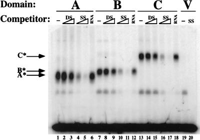
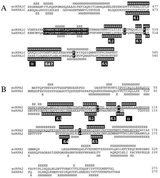
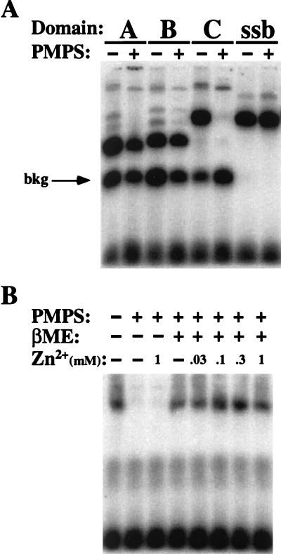
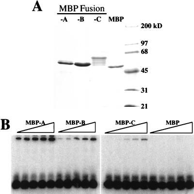
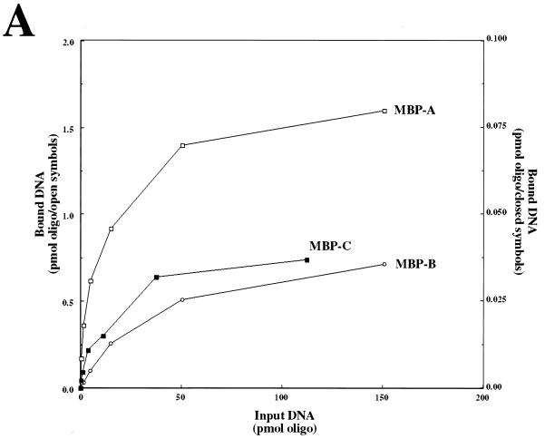
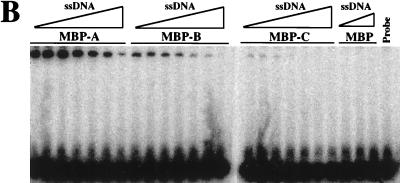
Replication protein A (RPA), the heterotrimeric single-stranded-DNA (ssDNA) binding protein (SSB) of eukaryotes, contains two homologous ssDNA binding domains (A and B) in its largest subunit, RPA1, and a third domain in its second-largest subunit, RPA2. Here we report that Saccharomyces cerevisiae RPA1 contains a previously undetected ssDNA binding domain (domain C) lying in tandem with domains A and B. The carboxy-terminal portion of domain C shows sequence similarity to domains A and B and to the region of RPA2 that binds ssDNA (domain D). The aromatic residues in domains A and B that are known to stack with the ssDNA bases are conserved in domain C, and as in domain A, one of these is required for viability in yeast. Interestingly, the amino-terminal portion of domain C contains a putative Cys4-type zinc-binding motif similar to that of another prokaryotic SSB, T4 gp32. We demonstrate that the ssDNA binding activity of domain C is uniquely sensitive to cysteine modification but that, as with gp32, ssDNA binding is not strictly dependent on zinc. The RPA heterotrimer is thus composed of at least four ssDNA binding domains and exhibits features of both bacterial and phage SSBs.


Figures

FIG. 1
FIG. 1
Identification of a new ssDNA binding domain in RPA1. Fragments of the scRPA1 protein were expressed in E. coli and tested for ssDNA binding activity by the UV EMSA. At the top is a schematic of the scRPA1 protein illustrating the locations of the three ssDNA binding domains and the eight protein fragments (numbered horizontal black bars) that were tested in this assay. Numbers above the schematic refer to the amino acid numbers of the scRPA1 protein. For each protein fragment, a portion of extract containing approximately 0.3 μg of recombinant protein was incubated with a 17-nt oligonucleotide 32P labeled at its 5′ end (P) and cross-linked with UV light. Products were resolved on a 6% denaturing polyacrylamide gel. V, extract made from E. coli expressing the vector alone; A*, B*, and C*, RPA1 domains A, B, and C; bkg, background band.
FIG. 2
FIG. 2
RPA1 domain C binds ssDNA specifically. Extracts of E. coli containing approximately 0.3 μg of recombinant protein were assayed in the presence of the indicated competitor, namely, 0.5 or 3 μg of unlabled lambda DNA before (double stranded [DS]) or after (single stranded [SS]) boiling or 3 μg of yeast tRNA (RNA). V, extract of E. coli expressing the vector alone or competed with 3 μg of ssDNA. A*, B*, and C*, RPA1 domains A, B, and C.
FIG. 3
FIG. 3
Amino acid sequence alignment of the four ssDNA binding domains of RPA. The amino acid sequences of the indicated proteins were aligned with the PILEUP program and optimized as described in Materials and Methods. The secondary structure of hsRPA1181–422, presented at the top of the sequences, was taken from Bochkarev et al. (5). Highly conserved residues are indicated in white type on a black background, and the putative zinc-binding domain in domain C is shaded. Numbers to the right of the protein designations refer to amino acid numbers. Note that 14 amino acids are deleted from the scRPA2 sequence at residue 115. Hs, H. sapiens; Xl, Xenopus laevis; Sc, S. cerevisiae; Sp, Schizosaccharomyces pombe; Os, Oryza sativa; Cf, Crithidia fasiculata; Ce, Caenorhabditis elegans.
FIG. 4
FIG. 4
Secondary-structure predictions of domains C and D are consistent with an OB fold. Amino acid sequences of domains C and D from yeast and humans were analyzed by the Predict Protein program, European Molecular Biology Laboratory, Heidelberg, Germany (48). The predicted secondary structures of domains C (A) and D (B) are displayed above the yeast (scRPA1C and scRPA2, respectively) and below the human (hsRPA1C and hsRPA2, respectively) sequences. The sequence of the Cys4 motif (A) and the three highly conserved residues within each amino acid sequence (A and B) are highlighted in white type on a black background. The proposed β strands of the OB fold are indicated. H, helix; E, extended β-sheet.
FIG. 5
FIG. 5
ssDNA binding activity of domain C requires the Cys4 motif. Extracts of E. coli expressing domain C of scRPA1 with the indicated mutations were assayed by UV EMSA. Vector is an extract of E. coli expressing the vector alone. ΔC20 is domain C with a C-terminal truncation of 20 amino acids. WT, wild type.
FIG. 6
FIG. 6
Domain C binding activity is sensitive to cysteine modification. (A) Extracts of E. coli expressing the indicated domains of scRPA1 or purified Ecssb (ssb) were incubated in the presence or absence of 2.5 mM PMPS for 1 h, made 50 mM in EDTA, and assayed for ssDNA binding activity by UV EMSA. (B) An extract of E. coli expressing domain C was treated with 2.5 mM PMPS for 1 h, made 50 mM in EDTA, and dialyzed against Chelex-100-treated TNG buffer containing 1 mM EDTA. The modified protein was then treated with the indicated reagents for 30 min before assay by UV EMSA. βME, β-mercaptoethanol; bkg, background.
FIG. 7
FIG. 7
ssDNA binding activity of MBP-RPA fusion proteins. (A) Two micrograms of each purified MBP fusion protein was resolved by SDS–10% PAGE and stained with Coommassie blue. (B) Increasing amounts of the indicated MBP fusion protein or native MBP were incubated with a fixed amount of probe, UV cross-linked, and resolved by denaturing gel electrophoresis. Protein titrations are 0.1, 0.3, 1, 3, and 10 μg in 15-μl binding reaction mixtures.
FIG. 8
FIG. 8
ssDNA binding activities of MBP-RPA fusion proteins are saturable and reversible. (A) Increasing amounts of input DNA were incubated with 0.3 μg of MBP-A (5.4 pmol), 0.3 μg of MBP-B (5.4 pmol), or 1 μg of MBP-C (15 pmol) and assayed by UV EMSA. MPB-C values correspond to the 1/20th scale on the right. oligo, oligonucleotide. (B) One microgram of each MBP fusion protein was preincubated with probe (10 fmol) and then incubated with 0, 1, 3, 10, 30, 100, and 300 pmol of unlabeled oligonucleotide. MBP alone was incubated with 0, 100, and 300 pmol unlabeled oligonucleotide. The last lane is a control lacking protein.
FIG. 8
FIG. 8
ssDNA binding activities of MBP-RPA fusion proteins are saturable and reversible. (A) Increasing amounts of input DNA were incubated with 0.3 μg of MBP-A (5.4 pmol), 0.3 μg of MBP-B (5.4 pmol), or 1 μg of MBP-C (15 pmol) and assayed by UV EMSA. MPB-C values correspond to the 1/20th scale on the right. oligo, oligonucleotide. (B) One microgram of each MBP fusion protein was preincubated with probe (10 fmol) and then incubated with 0, 1, 3, 10, 30, 100, and 300 pmol of unlabeled oligonucleotide. MBP alone was incubated with 0, 100, and 300 pmol unlabeled oligonucleotide. The last lane is a control lacking protein.

References

    1. Alani E, Thresher R, Griffith J D, Kolodner R D. Characterization of DNA-binding and strand-exchange stimulation properties of y-RPA, a yeast single-strand-DNA-binding protein. J Mol Biol. 1992;227:54–71. - PubMed
    1. Atrazhev A, Zhang S, Grosse F. Single-stranded DNA binding protein from calf thymus. Purification, properties, and stimulation of the homologous DNA-polymerase-alpha-primase complex. Eur J Biochem. 1992;210:855–865. - PubMed
    1. Blackwell L J, Borowiec J A. Human replication protein A binds single-stranded DNA in two distinct complexes. Mol Cell Biol. 1994;14:3993–4001. - PMC - PubMed
    1. Blackwell L J, Borowiec J A, Mastrangelo I A. Single-stranded-DNA binding alters human replication protein A structure and facilitates interaction with DNA-dependent protein kinase. Mol Cell Biol. 1996;19:4798–4807. - PMC - PubMed
    1. Bochkarev A, Pfuetzner R A, Edwards A M, Frappier L. Structure of the single-stranded-DNA-binding domain of replication protein A bound to DNA. Nature. 1997;385:176–181. - PubMed

Publication types

LinkOut - more resources