. 1998 Dec;18(12):7269-77.
doi: 10.1128/MCB.18.12.7269.

C/EBPalpha is critical for the neonatal acute-phase response to inflammation

Affiliations

C/EBPalpha is critical for the neonatal acute-phase response to inflammation

B L Burgess-Beusse et al. Mol Cell Biol. 1998 Dec.

Abstract

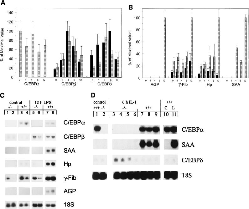
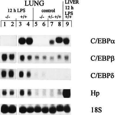
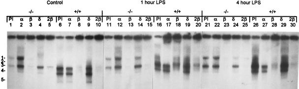
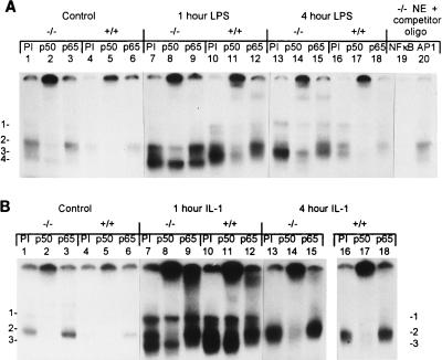
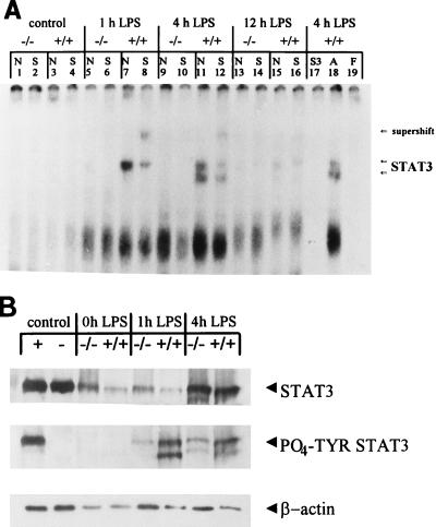
Members of the C/EBP (CCAAT/enhancer binding protein) family of transcription factors play important roles in mediating the acute-phase response (APR), an inflammatory process resulting from infection and/or tissue damage. Among the C/EBP family of proteins, C/EBPbeta and -delta were thought to be the primary mediators of the APR. The function of C/EBPalpha in the APR has not been fully characterized to date. Here, we investigate the role of C/EBPalpha in the APR by using neonatal mice that lack C/EBPalpha expression. Northern blot analysis of acute-phase protein gene expression in neonatal mice treated with purified bacterial lipopolysaccharide or recombinant interleukin 1beta as an inflammation stimulus showed a strong APR in wild-type mice, but a response in C/EBPalpha null animals was completely lacking. The C/EBPalpha knockout and wild-type mice demonstrated elevations in C/EBPbeta and -delta mRNA expression and DNA binding as well as increased DNA binding of NF-kappaB, all of which are known to be important in the APR. Null mice, however, failed to activate STAT3 binding in response to lipopolysaccharide. Our results provide the first evidence that C/EBPalpha is absolutely required for the APR in neonatal mice, is involved in STAT3 regulation, and cannot be compensated for by other C/EBP family members.


Figures

FIG. 1
FIG. 1
Lack of APP gene induction in C/EBPα−/− mice in response to LPS or IL-1. Northern analysis of 25 μg of total RNA from newborn C/EBPα−/− (−/−) and C/EBPα+/+ (+/+) mice at 0, 1, 4, 6, and 12 h post-LPS treatment. (A and B) Expression of C/EBP gene family (A) and APP genes (B). Blots were quantified, and the phosphorimaging value of each probe was normalized to the value of the 18S rRNA probe used as a loading control. The mean value for each genotype at each time point was determined. The largest normalized value for a given gene probe was set at 100%, and the normalized values of the other samples were compared to this maximal value. For each probe (indicated below the appropriate LPS time course), the data was expressed as the percentage of the maximal value ± standard deviation obtained for that probe. Each bar represents the results for three animals, except those for null and wild-type mice at 12 h post-LPS treatment (n = 5 and 4, respectively) and for null mice at 1 h post-LPS treatment (n = 4). ■, null mice; ▤, wild-type mice. (C) Representative Northern blots showing results for RNA from the same blot for two control (glucose-treated) −/− (lanes 1 and 2) and +/+ (lanes 3 and 4) pups and for RNA from the same blot for two −/− (lanes 5 and 6) and +/+ (lanes 7 and 8) mice at 12 h post-LPS treatment. The blots were hybridized with each individual probe sequentially, with the probe used indicated to the right of the panel. (D) Northern blot results for 25 μg of total liver RNA from control (glucose-treated) −/− (lane 2) and +/+ (lane 1) mice, 6-h-IL-1-treated −/− (lanes 3 to 6) and +/+ (lanes 7 to 9) animals, and +/+ control (C) (lane 10) and 6-h-LPS-treated (L) (lane 11) mice, hybridized sequentially with the probes indicated to the right of the panel.
FIG. 2
FIG. 2
LPS response in neonatal C/EBPα−/− mouse lung. Northern blot analysis of 25 μg of total lung RNA from 12-h-LPS-treated C/EBPα−/− (−/−) (lanes 1 and 2) and C/EBPα+/+ (+/+) (lanes 3 and 4) mice and control (glucose-treated) −/− (lanes 5 and 6), +/− (lane 7), and +/+ (lane 8) mice, with results for 25 μg of RNA from 12-h-LPS-treated mouse liver (lane 9) shown for comparison, hybridized sequentially with the indicated probes.
FIG. 3
FIG. 3
C/EBPβ and -δ are present in C/EBPα−/− mice and respond to LPS treatment. EMSA of C/EBP binding activity in C/EBPα−/− (−/−) and C/EBPα+/+ (+/+) mice treated with glucose (control) (lanes 1 to 10) or LPS for 1 (lanes 11 to 20) or 4 (lanes 21 to 30) h are shown. Complexes are numbered to the left of the figure. Ten micrograms of liver NE was preincubated with 1 μl of antisera against C/EBPα (α), C/EBPβ (β), C/EBPδ (δ), or PI serum as a control or with 2 μl of C/EBPβ (2β) antiserum to determine the nature of the complexes. The C/EBPα and C/EBPδ antisera showed a stabilizing effect on the NE proteins. The antisera used are indicated above the lanes.
FIG. 4
FIG. 4
Induction of NF-κB family members by LPS and IL-1 in all genotypes. EMSA analysis of NF-κB binding activity on 10 μg of liver NE from C/EBPα−/− (−/−) and C/EBPα+/+ (+/+) mice, with preincubation with 1 μl of PI serum used as a control, or of NF-κB p50 or p65 antisera to identify proteins in the complexes is shown. Genotypes and treatments are indicated above the lanes. The antiserum used is listed above each lane, and complexes are numbered on the left (A and B) and/or right (B) of the panel. (A) NF-κB response to LPS. Mice were treated with glucose (control) (lanes 1 to 6) or LPS for 1 (lanes 7 to 12) or 4 (lanes 13 to 18) h and analyzed for protein binding. Competition using 100-fold excess of either unlabeled NF-κB or AP1 (from Santa Cruz Biotechnology, Inc.) oligonucleotides (oligo) and 10 μg of liver NE from C/EBPα−/− mice demonstrated that the complexes were specific to NF-κB; the competitor is specified above the lanes (lanes 19 and 20). (B) NF-κB response to IL-1. Mice were treated with glucose (control) or IL-1 for 1 or 4 h and analyzed for protein binding. Samples in lanes 16 to 18 were run on a separate gel.
FIG. 5
FIG. 5
No STAT3 binding in response to LPS treatment in C/EBPα−/− mouse livers. (A) EMSA analysis of STAT3 binding activity by using 10 μg of liver NE from C/EBPα−/− (−/−) and C/EBPα+/+ (+/+) mice treated with glucose (control) (lanes 1 to 4) or LPS for 1 (lanes 5 to 8), 4 (lanes 9 to 12), or 12 (lanes 13 to 16) h, with preincubation with either no antiserum (N) or 1 μl of STAT3 antiserum (S) to identify proteins in the complexes. Genotypes and treatments are indicated above the lanes. The antisera used are listed above the lanes, and the positions of STAT3-containing complexes and antibody supershift are shown to the right of the panel. Competition using a 100-fold excess of either unlabeled STAT3 (S3) or AP1 (A) (from Santa Cruz Biotechnology, Inc.) oligonucleotides and 10 μg of liver NE from 4-h LPS-treated +/+ mice demonstrated that the complexes were specific to STAT3 (competitors are specified above the lanes [lanes 17 and 18]). Free probe (F) was run without proteins present (lane 19). (B) Western blot analysis of STAT3 protein levels in 40 μg of liver NE. The mouse genotypes and treatments are indicated above the lanes, and the antisera used as probes are indicated on the right, with the polyclonal STAT3 antibodies denoted as STAT3 and the tyrosine-phosphorylated STAT3 antibodies designated PO4-TYR STAT3. “Control” refers to the NIH 3T3 cell extracts (New England Biolabs) that were used to control for the position of STAT3 and the phosphorylation status of STAT3. The positive control extracts (+) contain tyrosine-phosphorylated STAT3, while the negative control extracts (−) contain nonphosphorylated STAT3. The untreated control mouse liver extracts are designated as 0h LPS.

References

    1. Akira S, Isshiki H, Sugita T, Tanabe O, Kinoshita S, Nishio Y, Nakajima T, Hirano T, Kishimoto T. A nuclear factor for IL-6 expression (NF-IL6) is a member of a C/EBP family. EMBO J. 1990;9:1897–1906. - PMC - PubMed
    1. Akira S, Kishimoto T. IL-6 and NF-IL6 in acute-phase response and viral infection. Immunol Rev. 1992;127:25–50. - PubMed
    1. Alam T, An M R, Mifflin R C, Hsieh C C, Ge X, Papaconstantinou J. Trans-activation of the alpha 1-acid glycoprotein gene acute phase responsive element by multiple isoforms of C/EBP and glucocorticoid receptor. J Biol Chem. 1993;268:15681–15688. - PubMed
    1. Alam T, An M R, Papaconstantinou J. Differential expression of three C/EBP isoforms in multiple tissues during the acute phase response. J Biol Chem. 1992;267:5021–5024. - PubMed
    1. Allen M C. An overview of long-term outcome. In: Witter F R, Keith L G, editors. Textbook of prematurity: antecedents, treatment, and outcome. Boston, Mass: Little, Brown & Co.; 1993. pp. 371–384.

Publication types

MeSH terms

LinkOut - more resources