. 1998 Dec;18(12):7432-43.
doi: 10.1128/MCB.18.12.7432.

The leukemic protein core binding factor beta (CBFbeta)-smooth-muscle myosin heavy chain sequesters CBFalpha2 into cytoskeletal filaments and aggregates

Affiliations

The leukemic protein core binding factor beta (CBFbeta)-smooth-muscle myosin heavy chain sequesters CBFalpha2 into cytoskeletal filaments and aggregates

N Adya et al. Mol Cell Biol. 1998 Dec.

Abstract

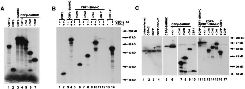
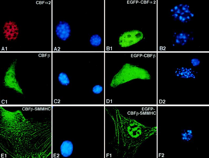
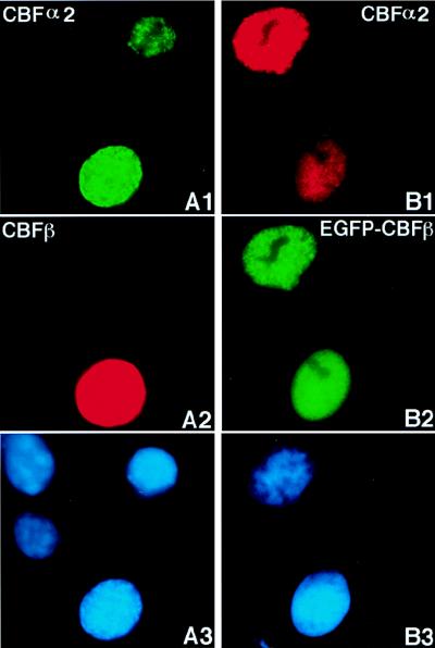
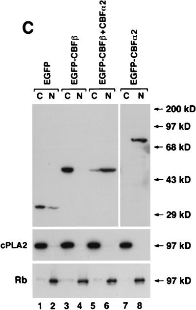
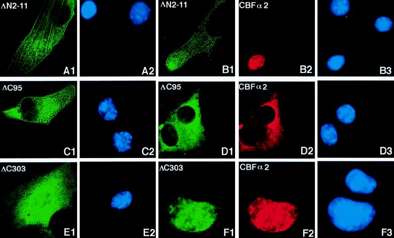
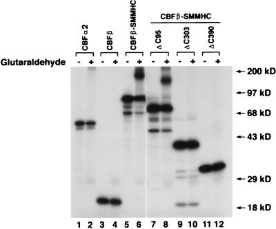
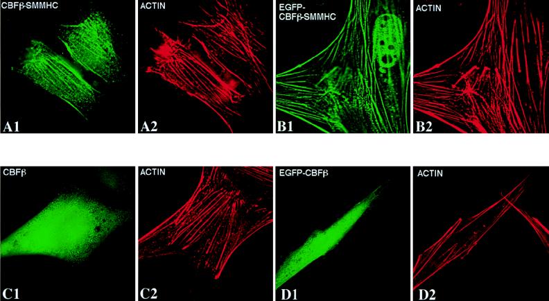
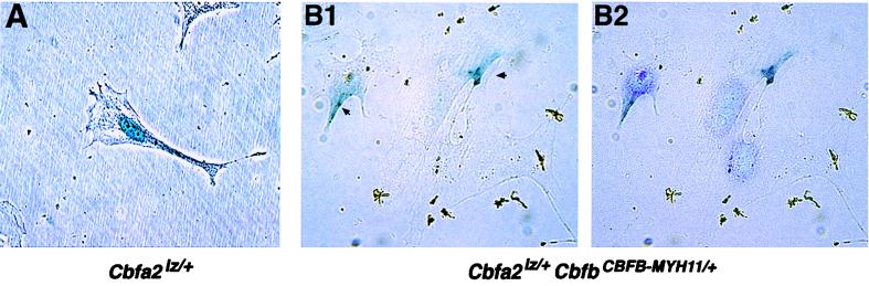
The fusion gene CBFB-MYH11 is generated by the chromosome 16 inversion associated with acute myeloid leukemias. This gene encodes a chimeric protein involving the core binding factor beta (CBFbeta) and the smooth-muscle myosin heavy chain (SMMHC). Mouse model studies suggest that this chimeric protein CBFbeta-SMMHC dominantly suppresses the function of CBF, a heterodimeric transcription factor composed of DNA binding subunits (CBFalpha1 to 3) and a non-DNA binding subunit (CBFbeta). This dominant suppression results in the blockage of hematopoiesis in mice and presumably contributes to leukemogenesis. We used transient-transfection assays, in combination with immunofluorescence and green fluorescent protein-tagged proteins, to monitor subcellular localization of CBFbeta-SMMHC, CBFbeta, and CBFalpha2 (also known as AML1 or PEBP2alphaB). When expressed individually, CBFalpha2 was located in the nuclei of transfected cells, whereas CBFbeta was distributed throughout the cell. On the other hand, CBFbeta-SMMHC formed filament-like structures that colocalized with actin filaments. Upon cotransfection, CBFalpha2 was able to drive localization of CBFbeta into the nucleus in a dose-dependent manner. In contrast, CBFalpha2 colocalized with CBFbeta-SMMHC along the filaments instead of localizing to the nucleus. Deletion of the CBFalpha-interacting domain within CBFbeta-SMMHC abolished this CBFalpha2 sequestration, whereas truncation of the C-terminal-end SMMHC domain led to nuclear localization of CBFbeta-SMMHC when coexpressed with CBFalpha2. CBFalpha2 sequestration by CBFbeta-SMMHC was further confirmed in vivo in a knock-in mouse model. These observations suggest that CBFbeta-SMMHC plays a dominant negative role by sequestering CBFalpha2 into cytoskeletal filaments and aggregates, thereby disrupting CBFalpha2-mediated regulation of gene expression.


Figures

FIG. 1
FIG. 1
Characterization of CBFα2, CBFβ, CBFβ-SMMHC, and the deletion mutants. (A) Generation of protein products by in vitro translation. [35S]methionine-labeled proteins were produced by in vitro translation, separated by gel electrophoresis, and detected by autoradiography. The intense bands at the bottom of the gel are the free [35S]methionine. (B) Interactions between CBFα2 and the deletion mutants. The protein products shown in panel A were immunoprecipitated with polyclonal antibody α3043, in the presence or absence of cold in vitro-translated CBFα2. (C) Expression of cDNA constructs after transient transfection into NIH 3T3 cells. The Western blot with extracts derived from transiently transfected NIH 3T3 cells was probed with α3043 to detect CBFα2 and EGFP-CBFα2 (lanes 1 to 4). The relevant proteins are shown by arrowheads in lanes 2 and 4. Similarly, the antibody β141.2 was used to identify CBFβ, CBFβ-SMMHC, the deletion proteins, and their GFP fusions by Western blotting of cellular extracts (lanes 5 to 17). Protein molecular mass markers are shown on the right sides of panels B and C.
FIG. 2
FIG. 2
Subcellular localization of CBFα2, CBFβ, and CBFβ-SMMHC in transiently transfected cells. NIH 3T3 cells were transfected with pCbfa2 (A1 and A2), pEGFP-Cbfa2 (B1 and B2), pCBFB (C1 and C2), pEGFP-CBFB (D1 and D2), pCBFB-MYH11 (E1 and E2), and pEGFP-CBFB-MYH11 (F1 and F2). Odd-numbered panels show fluorescent signals, while even-numbered panels show DAPI staining of the same fields. Panel A1 shows signal detected with α3043 and a Texas-red-labeled secondary antibody, panels C1 and E1 show signals detected with β141.2 and a fluorescein-labeled secondary antibody, and panels B1, D1, and F1 show the autofluorescent signal of GFP fused to the respective proteins. The antibodies failed to detect the endogenous proteins (compare the untransfected cell in panel A1 with panel A2, as well as that in panel C1 with panel C2).
FIG. 3
FIG. 3
CBFα2 brings CBFβ into the nucleus when coexpressed. Cells were cotransfected with pCbfa2 and pCBFB (A1, A2, and A3) or with pCbfa2 and pEGFP-CBFB (B1, B2, and B3). Panels A1, A2, and A3 represent images of the same field seen through different filters, as do panels B1, B2, and B3. CBFα2 was detected with antibody α3043 and a fluorescein-labeled (A1) or a Texas-red-labeled (B1) secondary antibody. CBFβ localization was detected with β141.2 and a Texas-red-labeled secondary antibody (A2). Panel B2 shows the autofluorescence of EGFP-CBFβ. Cells in panels A3 and B3 were stained by DAPI. (C) Cytoplasmic and nuclear fractionation of the transfected cells is shown. NIH 3T3 cells transfected with pEGFP (lanes 1 and 2), pEGFP-CBFB (lanes 3 and 4), pEGFP-CBFB plus pCbfa2 (lanes 5 and 6), and pEGFP-Cbfa2 alone (lanes 7 and 8) were fractionated into cytoplasmic and nuclear fractions, which were separated on SDS-PAGE gel and probed by Western blotting with a GFP-specific antibody. The quality of cell fractionation was analyzed by probing with antibodies for cPLA2 (cytoplasmic) and Rb (nuclear). C, cytoplasmic fraction; N, nuclear fraction. Molecular mass markers are shown to the right in panel C.
FIG. 3
FIG. 3
CBFα2 brings CBFβ into the nucleus when coexpressed. Cells were cotransfected with pCbfa2 and pCBFB (A1, A2, and A3) or with pCbfa2 and pEGFP-CBFB (B1, B2, and B3). Panels A1, A2, and A3 represent images of the same field seen through different filters, as do panels B1, B2, and B3. CBFα2 was detected with antibody α3043 and a fluorescein-labeled (A1) or a Texas-red-labeled (B1) secondary antibody. CBFβ localization was detected with β141.2 and a Texas-red-labeled secondary antibody (A2). Panel B2 shows the autofluorescence of EGFP-CBFβ. Cells in panels A3 and B3 were stained by DAPI. (C) Cytoplasmic and nuclear fractionation of the transfected cells is shown. NIH 3T3 cells transfected with pEGFP (lanes 1 and 2), pEGFP-CBFB (lanes 3 and 4), pEGFP-CBFB plus pCbfa2 (lanes 5 and 6), and pEGFP-Cbfa2 alone (lanes 7 and 8) were fractionated into cytoplasmic and nuclear fractions, which were separated on SDS-PAGE gel and probed by Western blotting with a GFP-specific antibody. The quality of cell fractionation was analyzed by probing with antibodies for cPLA2 (cytoplasmic) and Rb (nuclear). C, cytoplasmic fraction; N, nuclear fraction. Molecular mass markers are shown to the right in panel C.
FIG. 4
FIG. 4
CBFβ-SMMHC sequesters CBFα2 along filaments and aggregates. (A1, A2, and A3) NIH 3T3 cells were cotransfected with pCbfa2 and pCBFB-MYH11. CBFβ-SMMHC localization was detected with antibody β141.2 and a fluorescein-labeled secondary antibody (panel A1), and CBFα2 localization was detected with antibody α3043 and a Texas-red-labeled secondary antibody (panel A2). (B1, B2, and B3) Localization of EGFP-CBFα2 and CBFβ-SMMHC when cotransfected. CBFβ-SMMHC localization is shown by the red signal (β141.2 and a Texas-red-labeled secondary antibody) in panel B1, while EGFP-CBFα2 localization is shown by the green autofluorescent signal in panel B2. (C1, C2, and C3) Cotransfected EGFP-CBFβ-SMMHC (panel C1) and CBFα2 (panel C2), which were detected in a fashion similar to that described for panel B. (A3, B3, and C3) DAPI staining of the respective fields.
FIG. 5
FIG. 5
Immunofluorescence of CBFβ-SMMHC deletion proteins and coexpressed CBFα2. CBFβ-SMMHC deletion proteins and CBFα2 were detected with β141.2 and α3043, respectively, plus appropriate secondary antibodies. (A and B) Immunofluorescence labeling of NIH 3T3 cells transfected with pCBFB-MYH11ΔN2-11, either alone (panels A1 and A2) or with pCbfa2 (panels B1, B2, and B3). Panels A1 and A2 show the same field with the CBFβ-SMMHCΔN2-11 localization in green and DAPI-stained nuclei in blue, respectively. Localization of CBFβ-SMMHCΔN2-11 is again shown in green in panel B1, while cotransfected CBFα2 is shown in red in panel B2. (C1 and C2) Immunofluorescence labeling of cells transfected with only pCBFB-MYH11ΔC95. (D) Cells cotransfected with pCBFB-MYH11ΔC95 (panel D1) and pCbfa2 (panel D2). CBFβ-SMMHCΔC303 was labeled in green when transfected alone (E1) or cotransfected (F1) with CBFα2, which was labeled in red (F2). The panels labeled with the same letter are views of the same fields. (A2, B3, C2, D3, E2, and F3) DAPI staining showing the nuclei of cells in the respective fields.
FIG. 6
FIG. 6
Glutaraldehyde cross-linking of CBFβ, CBFβ-SMMHC, and the deletion proteins. Proteins as labeled at the top of the lanes were translated in vitro in the presence of [35S]methionine. Cross-linking of each protein was accomplished by incubation in the presence (even-numbered lanes) or absence (odd-numbered lanes) of glutaraldehyde. Slow mobility bands were observed for CBFβ-SMMHC and CBFβ-SMMHCΔC95 but not for CBFα2, CBFβ, CBFβ-SMMHCΔC303, and CBFβ-SMMHCΔC390. Protein molecular mass markers are shown on the right.
FIG. 7
FIG. 7
CBFβ-SMMHC colocalizes with actin filaments, while CBFβ does not. NIH 3T3 cells were transfected with pCBFB-MYH11 (A1 and A2), pEGFP-CBFB-MYH11 (B1 and B2), pCBFB (C1 and C2), and pEGFP-CBFB (D1 and D2). Proteins were detected with β141.2 plus a fluorescein isothiocyanate-labeled secondary antibody (panels A1 and C1) and Texas-red-conjugated phalloidin (panels A2, B2, C2, and D2). Panels B1 and D1 show the autofluorescent signal from the GFP fusion proteins.
FIG. 8
FIG. 8
In vivo sequestration of Cbfα2 by Cbfβ-SMMHC. Embryonic fibroblasts were isolated from 11.5-dpc mouse embryos, cultured briefly in vitro, and stained for the Cbfα2-βgal protein. (A) A fibroblast from an embryo of Cbfa2lz/+ genotype stained for βgal. (B1) Fibroblasts from an embryo of Cbfa2lz, CbfbCBFB-MYH11/+ genotype stained for βgal. Arrows indicate the βgal staining. (B2) DAPI staining of the same field as shown in panel B1.

References

    1. Adya, N., and P. P. Liu. Unpublished data.
    1. Arthur D C, Bloomfield C D. Association of partial deletion of the long arm of chromosome 16 and bone marrow eosinophilia in acute non-lymphocytic leukemia. Blood. 1983;62:931. . (Letter.) - PubMed
    1. Bae S-C, Ogawa E, Maruyama M, Oka H, Satake M, Shigesada K, Jenkins N A, Gilbert D J, Copeland N G, Ito Y. PEBP2αB/mouse AML1 consists of multiple isoforms that possess differential transactivation potentials. Mol Cell Biol. 1994;14:3242–3252. - PMC - PubMed
    1. Bae S C, Yamaguchi-Iwai Y, Ogawa E, Maruyama M, Inuzuka M, Kagoshima H, Shigesada K, Satake M, Ito Y. Isolation of PEBP2 α B cDNA representing the mouse homolog of human acute myeloid leukemia gene, AML1. Oncogene. 1993;8:809–814. - PubMed
    1. Cameron S, Taylor D S, TePas E C, Speck N A, Mathey-Prevot B. Identification of a critical regulatory site in the human interleukin-3 promoter by in vivo footprinting. Blood. 1994;83:2851–2859. - PubMed

Publication types

MeSH terms

LinkOut - more resources