. 1998 Dec;18(12):7510-20.
doi: 10.1128/MCB.18.12.7510.

The pre-mRNA 5' cap determines whether U6 small nuclear RNA succeeds U1 small nuclear ribonucleoprotein particle at 5' splice sites

Affiliations

The pre-mRNA 5' cap determines whether U6 small nuclear RNA succeeds U1 small nuclear ribonucleoprotein particle at 5' splice sites

L O'Mullane et al. Mol Cell Biol. 1998 Dec.

Abstract

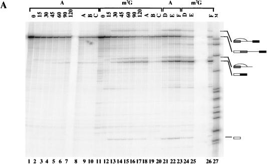
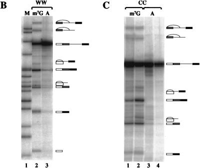
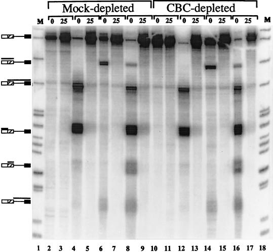
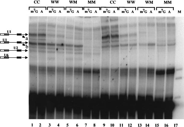
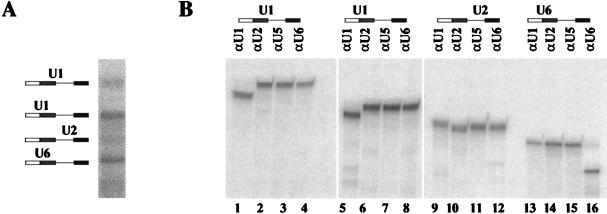
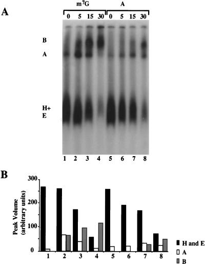
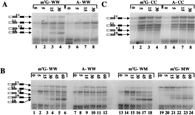
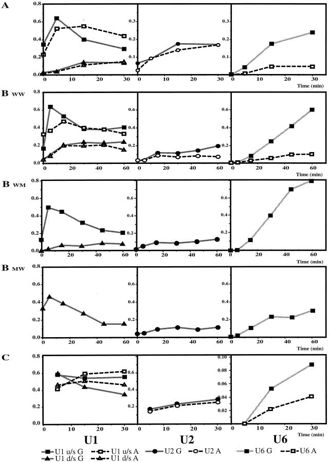
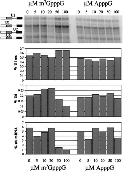
Efficient splicing of the 5'-most intron of pre-mRNA requires a 5' m7G(5')ppp(5')N cap, which has been implicated in U1 snRNP binding to 5' splice sites. We demonstrate that the cap alters the kinetic profile of U1 snRNP binding, but its major effect is on U6 snRNA binding. With two alternative wild-type splice sites in an adenovirus pre-mRNA, the cap selectively alters U1 snRNA binding at the site to which cap-independent U1 snRNP binding is stronger and that is used predominantly in splicing; with two consensus sites, the cap acts on both, even though one is substantially preferred for splicing. However, the most striking quantitative effect of the 5' cap is neither on U1 snRNP binding nor on the assembly of large complexes but on the replacement of U1 snRNP by U6 snRNA at the 5' splice site. Inhibition of splicing by a cap analogue is correlated with the loss of U6 interactions at the 5' splice site and not with any loss of U1 snRNP binding.


Figures

FIG. 1
FIG. 1
Pre-mRNA substrates containing alternative 5′ splice sites require the m7GpppG 5′ cap structure for splicing to both sites. (A) Splicing of ApppG- and m7GpppG-capped human β-globin IVS-1 substrate psp64 5′Δ16, which contains alternative 5′ splice sites. ApppG-capped RNA is in lanes 1 to 10 and 21 to 23; m7GpppG-capped RNA is in lanes 11 to 20 and 24 to 26. Splicing reaction mixtures were incubated for the times indicated (in minutes) above each lane. Lanes 8 to 10 and 18 to 26 were incubated for 120 min. Lanes A, B, and C contain 0, 100, and 1 mM m7GpppG cap analogue as a competitor. Lanes D, E, and F contain increasing amounts of SR proteins: 0.75, 1.5, and 3 μg. Cap analogues and SR proteins were preincubated with the extract before the addition of the pre-mRNA and splicing mix. Lane 27 (M) contains size markers. Spliced products and intermediates were resolved on a 6% denaturing polyacrylamide gel. (B) Splicing of m7GpppG- and ApppG-capped adenovirus pre-mRNA with a duplicated wild-type 5′ splice site (Ad1WW). (C) Splicing of adenovirus pre-mRNA with duplicated consensus 5′ splice sites (Ad1CC). Splicing reactions were for 40 min (lanes 1 and 3) or 1.5 h (lanes 2 and 4).
FIG. 1
FIG. 1
Pre-mRNA substrates containing alternative 5′ splice sites require the m7GpppG 5′ cap structure for splicing to both sites. (A) Splicing of ApppG- and m7GpppG-capped human β-globin IVS-1 substrate psp64 5′Δ16, which contains alternative 5′ splice sites. ApppG-capped RNA is in lanes 1 to 10 and 21 to 23; m7GpppG-capped RNA is in lanes 11 to 20 and 24 to 26. Splicing reaction mixtures were incubated for the times indicated (in minutes) above each lane. Lanes 8 to 10 and 18 to 26 were incubated for 120 min. Lanes A, B, and C contain 0, 100, and 1 mM m7GpppG cap analogue as a competitor. Lanes D, E, and F contain increasing amounts of SR proteins: 0.75, 1.5, and 3 μg. Cap analogues and SR proteins were preincubated with the extract before the addition of the pre-mRNA and splicing mix. Lane 27 (M) contains size markers. Spliced products and intermediates were resolved on a 6% denaturing polyacrylamide gel. (B) Splicing of m7GpppG- and ApppG-capped adenovirus pre-mRNA with a duplicated wild-type 5′ splice site (Ad1WW). (C) Splicing of adenovirus pre-mRNA with duplicated consensus 5′ splice sites (Ad1CC). Splicing reactions were for 40 min (lanes 1 and 3) or 1.5 h (lanes 2 and 4).
FIG. 2
FIG. 2
RNase H protection of consensus 5′ splice sites. The substrate pre-mRNA was m7GpppG-capped C174C, derived from β-globin IVS-2, which contained two alternative consensus 5′ splice sites. Pre-mRNA was incubated in HeLa nuclear extract that had been depleted of CBC or had been mock depleted. Oligonucleotides that direct RNase H cleavage to the consensus 5′ splice sites were added either just before the pre-mRNA (0) or 25 min after the pre-mRNA (25). No MgCl2 or ATP was added to the reaction mixtures. No oligonucleotide was added to the reaction mixtures in lanes 2, 3, 10, and 11; an oligonucleotide directed against the upstream site was added in lanes 4, 5, 12, and 13; an oligonucleotide directed against the downstream site was added in lanes 6, 7, 14, and 15; both were added to the reaction mixtures in lanes 8, 9, 16, and 17. The reaction mixtures were resolved on a 6% polyacrylamide gel. The products of RNase H cleavage are indicated with bars above the diagrams of the pre-mRNA on the left-hand side.
FIG. 3
FIG. 3
Psoralen cross-linking of U1 snRNA to m7GpppG- and ApppG-capped Ad1 substrates. The substrates used had two alternative 5′ splice sites, each of which was either consensus (C), wild-type (W), or mutant (M), as indicated above each lane. (A) Pre-mRNA capped with m7GpppG (m7G) or ApppG (A) was incubated for 20 min in splicing mixes containing AMT-psoralen in the presence (lanes 1 to 8) or absence (lanes 9 to 16) of magnesium, ATP, and phosphocreatine. Cross-linked products were resolved on a 5% polyacrylamide gel. The positions of the snRNA cross-links are shown diagramatically. Bands a and b are minor forms of the two U1 cross-links.
FIG. 4
FIG. 4
Identification of snRNA cross-linked to pre-mRNA. (A) Splicing reaction mixtures containing m7GpppG-capped Ad1WW were irradiated (as shown in Fig. 3, lane 3) and resolved on a 5% preparative polyacrylamide gel. (B) The cross-linked products shown in panel A were digested with RNase H and oligonucleotides complementary to either U1, U2, U5, or U6 snRNA and then fractionated onto a 5% polyacrylamide gel. The diagram above each gel indicates the identity of the snRNA shown to be cross-linked to the pre-mRNA.
FIG. 5
FIG. 5
(A) Native gel analysis of splicing complexes formed with m7GpppG and ApppG primed Ad1 pre-mRNA. The incubation times for the splicing reactions are shown. The presumed identities of the bands are shown as complexes B, A, and H plus E. (B) Quantitative analysis of the data in panel A. Numbers correspond to the lanes in panel A.
FIG. 6
FIG. 6
Effects of the 5′ cap and splice site sequence on snRNA interactions with pre-mRNA during time courses of splicing reactions. (A) Ad1WW pre-mRNA, capped with m7GpppG or ApppG, was incubated under splicing conditions in the presence of psoralen at 30°C. After the times shown above the lanes, reaction mixtures were removed and irradiated for 1 min each at ambient temperature. The results were analyzed by gel electrophoresis. The bands at the foot of the panel are intramolecular cross-links. Parallel reactions included (i) duplicates that contained an exogenous protein but showed the same features on quantitative analysis, (ii) reactions in the absence of MgCl2 and ATP, and (iii) reactions that were incubated for 40 and 90 min but not irradiated to allow splicing efficiency to be measured. (B) Ad1WW, Ad1WM, and Ad1MW pre-mRNAs were incubated under splicing conditions as in panel A, but for longer periods; irradiated; and analyzed as in panel A. The unlabelled lanes contained RNA from parallel reactions for 2.7 h that had not been irradiated so that the efficiency of splicing could be measured. The band shown as comprising U6 snRNA cross-linked to the downstream site, which is most obvious in the reaction of Ad1MW (lanes 21 to 24), has not been demonstrated directly to be this molecule, but its substrate specificity, kinetics, and mobility are consistent with this identification. (C) Identical to panel A in all respects, including the parallel reactions, but with Ad1CC pre-mRNA.
FIG. 7
FIG. 7
Quantitative analysis of the cross-links shown in Fig. 6A, B, and C refers to the three panels of Fig. 6, with the corresponding substrate designations for panel B. The y axis in every case shows the percentage of the cross-linked RNA molecule in the reaction mixture (see Materials and Methods); the x axis shows the time of incubation under splicing conditions (in minutes). The reactions of RNA capped with m7GpppG and ApppG are shown on the same graphs. The three graphs in each row show results for U1, U2 and U6 cross-links, respectively, with the identities of the lines shown below. u/s, upstream; d/s, downstream.
FIG. 8
FIG. 8
Inhibition of U6 snRNA interactions with m7GpppG-capped Ad1WW pre-mRNA by the m7GpppG cap analogue but not the ApppG cap analogue. HeLa nuclear extract was preincubated with the m7GpppG or the ApppG cap analogue for 15 min prior to the addition of the pre-mRNA. The final concentrations of m7GpppG and ApppG were 0, 5, 10, 20, 50, and 100 μM, as marked above the gel. The cross-linked RNA was resolved on a 5% polyacrylamide gel. The bar charts aligned below the image show the relative intensities of specific products and the pre-mRNA (expressed as percentages) for each lane. The products measured are U1 cross-linked to the upstream 5′ splice site (the major site of splicing), U6 cross-linked, and the upstream (u/s) mRNA, as indicated by the y axis labels.

References

    1. Berget S M. Exon recognition in vertebrate splicing. J Biol Chem. 1995;270:2411–2414. - PubMed
    1. Bindereif A, Green M R. An ordered pathway of snRNP binding during mammalian pre-messenger RNA splicing complex assembly. EMBO J. 1987;6:2415–2424. - PMC - PubMed
    1. Cao W H, Jamison S F, Garcia-Blanco M A. Both phosphorylation and dephosphorylation of ASF/SF2 are required for pre-mRNA splicing in vitro. RNA. 1997;3:1456–1467. - PMC - PubMed
    1. Chabot B. Directing alternative splicing: cast and scenarios. Trends Genet. 1996;12:472–477. - PubMed
    1. Chabot B, Steitz J A. Recognition of mutant and cryptic 5′ splice sites by the U1 small nuclear ribonucleoprotein in vitro. Mol Cell Biol. 1987;7:698–707. - PMC - PubMed

Publication types

LinkOut - more resources