. 1998 Dec 1;18(23):9594-600.
doi: 10.1523/JNEUROSCI.18-23-09594.1998.

Neuroprotection by glial metabotropic glutamate receptors is mediated by transforming growth factor-beta

Affiliations

Neuroprotection by glial metabotropic glutamate receptors is mediated by transforming growth factor-beta

V Bruno et al. J Neurosci. .

Abstract

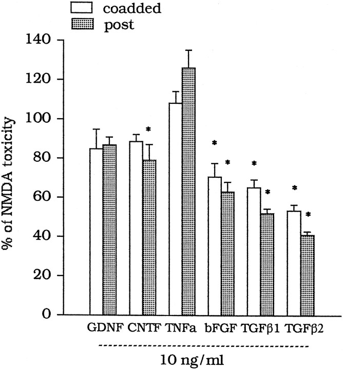
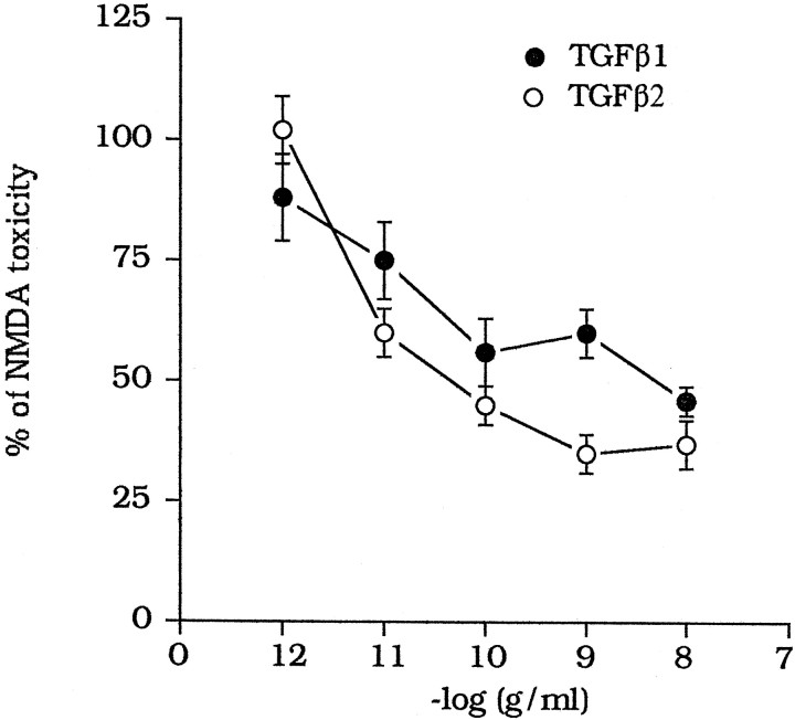
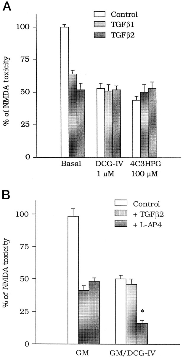
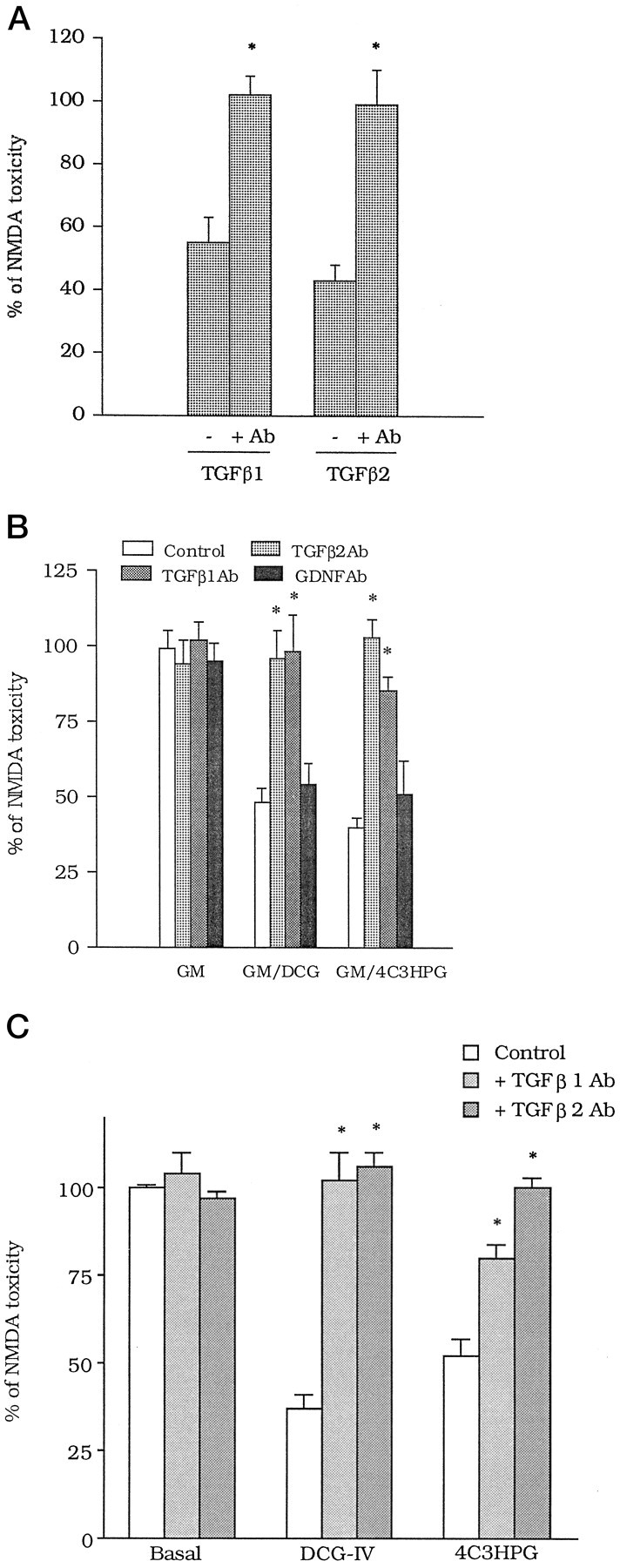
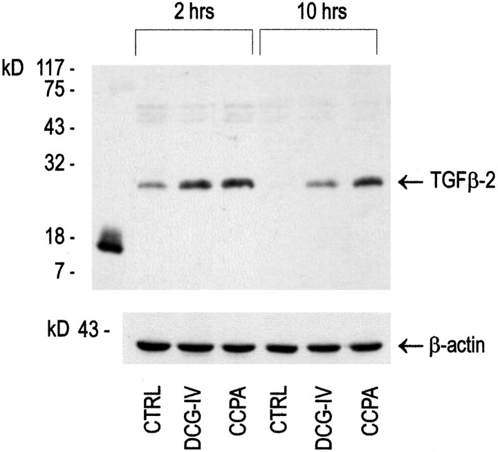
The medium collected from cultured astrocytes transiently exposed to the group-II metabotropic glutamate (mGlu) receptor agonists (2S,1'R, 2'R,3'R)-2-(2,3-dicarboxycyclopropyl)glycine (DCG-IV) or (S)-4-carboxy-3-hydroxyphenylglycine (4C3HPG) is neuroprotective when transferred to mixed cortical cultures challenged with NMDA (). The following data indicate that this particular form of neuroprotection is mediated by transforming growth factor-beta (TGFbeta). (1) TGFbeta1 and -beta2 were highly neuroprotective against NMDA toxicity, and their action was less than additive with that produced by the medium collected from astrocytes treated with DCG-IV or 4C3HPG (GM/DCG-IV or GM/4C3HPG); (2) antibodies that specifically neutralized the actions of TGFbeta1 or -beta2 prevented the neuroprotective activity of DCG-IV or 4C3HPG, as well as the activity of GM/DCG-IV or GM/4C3HPG; and (3) a transient exposure of cultured astrocytes to either DCG-IV or 4C3HPG led to a delayed increase in both intracellular and extracellular levels of TGFbeta. We therefore conclude that a transient activation of group-II mGlu receptors (presumably mGlu3 receptors) in astrocytes leads to an increased formation and release of TGFbeta, which in turn protects neighbor neurons against excitotoxic death. These results offer a new strategy for increasing the local production of neuroprotective factors in the CNS.


Figures

Fig. 1.
Fig. 1.
Effect of trophic factors on NMDA toxicity in mixed cultures of cortical cells. Factors were either combined with NMDA (coadded) or applied to the cultures immediately after the NMDA pulse and maintained in the medium during the following 20 hr (post). Values are means ± SEM of four to six determinations from two different multiplates and were calculated from the counts of neurons stained with trypan blue. Results were virtually identical when calculated from the extracellular LDH activity, which was always measured in parallel. In each multiplate, the mean of values obtained from individual dishes treated with NMDA alone, after subtracting the basal values, was considered as 100% or NMDA toxicity. Each individual determination was expressed as percentage of NMDA toxicity, always after subtracting the respective basal values. The SD calculated from the original counts of neurons stained with trypan blue was always <10% of the mean value for each experimental group. *p < 0.01 (one-way ANOVA + Fisher PLSD), if compared with values obtained with NMDA alone.
Fig. 2.
Fig. 2.
Concentration-dependent neuroprotection by TGFβ1 or -β2 against NMDA toxicity in mixed cultures of cortical cells. TGFβ1 or -β2 were applied immediately after the NMDA pulse and maintained in the medium during the following 20 hr. Values are means ± SEM of four individual determinations and were calculated from the counts of neurons stained with trypan blue, as described in Figure 1.
Fig. 3.
Fig. 3.
A, Lack of additive effects between TGFβ1 or -β2 and group-II mGlu receptor agonists on NMDA toxicity in mixed cortical cultures. DCG-IV (1 μm) or 4C3HPG (100 μm) were applied in combination with NMDA, whereas TGFβ1 or -β2 (both at 1 ng/ml) were applied after the NMDA pulse. Values are means ± SEM of four determinations. B, The neuroprotective activity of TGFβ2 (1 ng/ml) is obliterated when the factor is combined with the glial medium collected 20 hr after a 10 min exposure to 1 μm DCG-IV (GM/DCG-IV). Note that the protective activity of l-AP4 (100 μm) is instead additive to that produced by the medium of DCG-IV-treated astrocytes. GM, Glial medium. Values are means ± SEM of four determinations. *p < 0.01 (one-way ANOVA + Fisher PLSD) if compared with the respective controls. InA and B, values were calculated from the counts of neurons stained with trypan blue, as described in Figure1.
Fig. 4.
Fig. 4.
A, Antibodies (Ab) specific for TGFβ1 or -β2 (both at 100 ng/ml) abolish the neuroprotective activity of TGFβ1 and -β2 (both at 10 ng/ml) in mixed cortical cultures. Each of the factors and the respective antibody were added to the cultures immediately after the NMDA pulse and maintained in the medium during the following 20 hr. The concentration of antibodies was 100 ng/ml. Values are means ± SEM of eight determinations from two different experiments. *p < 0.01 (Student’s t test), if compared with the respective values obtained in the absence of the antibody. TGFβ1Ab at least did not prevent neuroprotection induced by bFGF applied after the NMDA pulse [bFGF, 10 ng/ml = 70 ± 3.5; bFGF + TGFβ1Ab (100 ng/ml) = 66 ± 4.2; TGFβ1Ab alone = 92 ± 6.3; n = 4, expressed as percentage of NMDA toxicity]. B, TGFβ1Ab and TGFβ2 Ab (both at 100 ng/ml) prevent the neuroprotective activity of the glial medium collected 20 hr after treating cultured astrocytes with 1 μm DCG-IV (GM/DCG-IV) or 100 μm 4C3HPG (GM/4C3HPG). Note that antibodies against GDNF (GDNFAb, 100 ng/ml) are inactive. All antibodies were applied to the glial medium before it was transferred to mixed cortical cultures. Values are means ± SEM of eight determinations from two different experiments; *p < 0.01 (one-way ANOVA + Fisher PLSD) if compared with the respective controls.C, TGFβ1Ab and TGFβ2Ab (all at 100 ng/ml) reduce the neuroprotective activity of group-II mGlu receptor agonists in mixed cortical cultures. DCG-IV (1 μm) or 4C3HPG (100 μm) were applied to the cultures during the 10 min pulse with NMDA. Antibodies were applied immediately after the pulse and maintained in the medium during the following 20 hr. Values are means ± SEM of four determinations. *p < 0.01 (one-way ANOVA + Fisher PLSD), if compared with the respective controls. In A–C, values were calculated from the counts of neurons stained with trypan blue, as described in Figure1.
Fig. 5.
Fig. 5.
Western blot analysis of TGFβ2 in protein extracts from cultured astrocytes transiently exposed to 1 μm DCG-IV or to 100 nm CCPA.CTRL, Control astrocyte cultures treated with buffer alone for 10 min, and then incubated for 2 or 10 hr in serum-free medium (see Materials and Methods); DCG-IV, cultures treated for 10 min with 1 μm DCG-IV and then incubated for 2 or 10 hr in serum-free medium; CCPA, cultures treated for 10 min with CCPA and then incubated for 2 or 10 hr in serum-free medium. Expression of β-actin is shown in the same protein extracts. Authentic monomeric human TGFβ2 is shown in thefirst lane.
Fig. 6.
Fig. 6.
A–F, Local infusion of TGFβ1 or -β2 protects against NMDA toxicity in the rat caudate nucleus. The core of the lesion in animals injected with NMDA, NMDA + TGFβ1, and NMDA + TGFβ2 is shown in A, B, andC, respectively. The white arrow points to a large area of necrosis, which is absent in B andC. Neuronal degeneration present at the periphery of the lesion in animals injected with NMDA is shown in D. The corresponding regions in animals injected with NMDA + TGFβ1 and NMDA + TGFβ2 are shown in E and F. The small areas indicated by the white arrowheads are also shown at higher magnification. Note the greater number of spared neurons in animals treated with TGFβ1 or -β2. G, Striatal GAD activity in animal locally infused with NMDA or with NMDA + TGFβ1 or NMDA + TGFβ2. Results are expressed as percentage of the respective contralateral unlesioned site for each individual determination. GAD activity was calculated as counts per minute of authentic [3H]GABA/μg of protein. *p< 0.05 (one-way ANOVA + Fisher PLSD) versus NMDA alone.

References

    1. Adams J, Collaco-Moraes Y, de Belleroche J. Cyclooxygenase-2 induction in cerebral cortex: an intracellular response to synaptic excitation. J Neurochem. 1996;66:6–13. - PubMed
    1. Bruno V, Sureda FX, Storto M, Casabona G, Caruso A, Knopfel T, Kuhn R, Nicoletti F. The neuroprotective activity of group-II metabotropic glutamate receptors requires new protein synthesis and involves a glial-neuronal interaction. J Neurosci. 1997;17:1891–1897. - PMC - PubMed
    1. Bruno V, Wroblewska B, Wroblewska JT, Fiore L, Nicoletti F. Neuroprotective activity of N-acetylaspartylglutamate in cultured cortical cells. Neuroscience. 1998;3:751–757. - PubMed
    1. Buisson A, Choi DW. The inhibitory mGluR agonist, S-4-carboxy-3-hydroxyphenylglycine, selectively attenuates NMDA neurotoxicity and oxygen-glucose deprivation-induced neuronal death. Neuropharmacology. 1995;34:1081–1087. - PubMed
    1. Buisson A, Nicole O, Nouvelot A, MacKenzie ET, Vivien D. Reduction of NMDA-induced toxicity by transforming growth factor-β1. Soc Neurosci Abstr. 1997;23:897.

MeSH terms

Substances

LinkOut - more resources