. 1998 Dec 1;18(23):9685-94.
doi: 10.1523/JNEUROSCI.18-23-09685.1998.

Axotomy reduces the effect of analgesic opioids yet increases the effect of nociceptin on dorsal root ganglion neurons

Affiliations

Axotomy reduces the effect of analgesic opioids yet increases the effect of nociceptin on dorsal root ganglion neurons

F A Abdulla et al. J Neurosci. .

Abstract

There is some doubt as to the effectiveness of opioids in the management of neuropathic pain. We therefore examined the actions of morphine and the opioid-like peptide nociceptin (both 1 mu) on dorsal root ganglion (DRG) neurons that were isolated from control or from nerve-injured rats. Both substances reduced omega-conotoxin (CTX) GVIA-sensitive, N-type Ca2+ channel current and small persistent nifedipine/ CTX-insensitive (non-N, non-L type) current. Nifedipine-sensitive L-type current was unaffected. The effect of nociceptin was antagonized by naloxone benzoylhydrazone (nalbzoh) but not by naloxone. Sciatic nerve section (axotomy) profoundly reduced the effects of morphine and the mu-receptor agonist D-ala2, N-Me-Phe4,Gly-ol5 enkephalin (DAMGO). The effect of the kappa-agonist [(+)-(5alpha,7alpha, 8beta)-N-methyl-N-(7-(1-pyrrolidinyl)-1-oxaspiro(4, 5)dec-8-yl)-benzeneacetamide] (U69593) was unchanged, whereas the effect of nociceptin was increased. All agonists produced their strongest effects on the small, putative nociceptive cells and their weakest effects on the largest cells. The delta-receptor agonist, enkephalin D-pen2,5 (DPDPE), was without effect on control or on axotomized cells. These and other data suggest that the functional downregulation of mu-opioid receptors on sensory nerves contributes to the poor efficacy of opioids in neuropathic pain. Also, the increased effectiveness of nociceptin after axotomy supports the hypothesis that its actions are mediated via a "non-opioid" receptor. Pronounced suppression of Ca2+ channel current in axotomized DRG neurons by nociceptin led to a reduction in Ca2+-dependent K+ conductance and a marked increase in excitability. Despite this, the spinal administration of nociceptin or agonists that activate ORL1 (opioid-like orphan receptor) may prove to be of clinical interest in the management of neuropathic pain.


Figures

Fig. 1.
Fig. 1.
Effects of morphine and nociceptin onIBa in DRG cells. A, Sample records from small cells from control (A1) and axotomized (A2) animals. Currents were recorded at −10 mV from a Vh of −90 mV. Tails were recorded at −40 mV, and voltage records were omitted for clarity. Both morphine and nociceptin decreased IBa in the cells illustrated both before and after axotomy. Note that theIBa suppression induced by 1 μm morphine was similar to that induced by nociceptin in the control cell, whereas in the axotomized cell the effect of morphine was decreased and that of nociceptin was increased. B, Time course of the effects of morphine or nociceptin on cells illustrated in A1 andA2 (data normalized). C, Graphs to show percentage suppression of IBain large (L), medium (M), and small (S) neurons from control or axotomized animals in response to 1 μm morphine or nociceptin. D, Graphs show percentage of cells in each category responding to 1 μmmorphine or to 1 μm nociceptin before or after axotomy.
Fig. 2.
Fig. 2.
Effects of μ-, κ-, and δ-agonists onIBa in control and axotomized DRG cells.A, Effects of the μ-agonist DAMGO and the κ-agonist U69593 and the lack of effect of the δ-agonist DPDPE (all 1 μm) on a small cell from a control animal.B, Effects of the same three agonists on a small cell from an axotomized animal. Note small response to DAMGO and lack of effect of DPDPE. The response to U69593 is similar to that seen in the control cell. Current and time calibrations refer to all records. Currents evoked at −10 mV from Vh = −90 mV. Each panel shows superimposed recordings ofIBa evoked before and during superfusion of drugs. Voltage command trace was omitted for clarity.
Fig. 3.
Fig. 3.
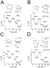
Effects of ω-CTX GVIA and nifedipine onIBa suppression induced by morphine and nociceptin. Superimposed current records in all parts of the figure areIBa responses to a step to −10 mV from aVh of −90 mV in the presence and absence of morphine or nociceptin (both 1 μm) and/or nifedipine (2 μm) or ω-CTX GVIA (1 μm). Time and current calibration in first record applies to all traces; voltage records were omitted for clarity. A, Left-hand data records show suppression of current by morphine. Right-hand data records comprise superimposed wash response (Wash) and current recorded in the presence of nifedipine and again in the presence of morphine plus nifedipine. The amount of IBa suppression produced is seen most clearly from the time course graph. This shows amplitudes of successive IBa responses recorded once every 20 sec in the presence of morphine, nifedipine, and nifedipine plus morphine. Nifedipine reduces the total current but does not impair the action of morphine. B, Left-hand data records are from another small cell, again showing suppression of current by morphine. Right-hand data records comprise superimposed wash response (Wash) and current recorded in the presence of ω-CTX GVIA and again in the presence of morphine plus ω-CTX GVIA. The time course graph shows that morphine is much less effective in attenuating the total IBa in the presence of ω-CTX GVIA. However, a clear reduction of the current is still observed.C, Data records and time course of an experiment performed with nociceptin and nifedipine. The ability of nociceptin to inhibit total IBa is not impaired by nifedipine. D, Data records and time course of an experiment performed with nociceptin and ω-CTX GVIA. Although the toxin impairs nociceptin-induced IBasuppression, the effect is not completely blocked.
Fig. 4.
Fig. 4.
Effects of morphine or nociceptin on the excitability of DRG cells. A, Sample records from a control small cell (A1) and an axotomized small cell (A2) illustrating the effects of 1 μm morphine and 1 μm nociceptin on excitability. To generate APs, both cells were depolarized with a 1 sec pulse of current at threshold strength (current trace omitted for clarity). Note that both morphine and nociceptin increase the excitability of the control cell in a similar way; however, in the axotomized cell, the response to morphine was much less than the response to nociceptin. B, Time course of the effects of morphine and nociceptin on cells illustrated in A1 and A2(data normalized). C, Graphs summarize effects of morphine or nociceptin on the percentage change in numbers of spikes discharged by 1 sec pulses of current at threshold strength in large (L), medium (M), and small (S) neurons from control or axotomized animals. D, Graphs show percentages of large, medium, and small neurons from control or axotomized animals that respond to morphine or nociceptin.
Fig. 5.
Fig. 5.
Effects of morphine or nociceptin (both 1 μm) on maximal outward current recorded at +70 mV (Vh = −90 mV). A, Superimposed data records of suppression of maximal outward current by 1 μm morphine or nociceptin in a control (A1) and in an axotomized (A2) small cell (voltage commands omitted for clarity). After initial suppression and recovery of current by morphine or nociceptin, the current is again suppressed after superfusion of 1 mm Cd2+ to blockgK, Ca responses. Note that in the control cell morphine and nociceptin produced similar effects on the outward current; however, after axotomy the effect of morphine was much smaller than that of nociceptin. Neither morphine nor nociceptin has effects on outward current recorded in the presence of Cd2+.B, Time course of the effects of morphine and nociceptin on cells illustrated in A1 andA2 (current is normalized for convenient comparison). C, Graphs summarizing effects of morphine and nociceptin (1 μm) on maximal outward current amplitude in large (L), medium (M), and small (S) neurons from control or axotomized animals. D, Graphs show percentages of large, medium, and small neurons from control or axotomized animals that respond to morphine or nociceptin (both 1 μm).

References

    1. Abdulla FA, Smith PA. Ectopic a2-adrenoceptors couple to N-type Ca2+ channels in axotomized rat sensory ganglia. J Neurosci. 1997a;17:1633–1641. - PMC - PubMed
    1. Abdulla FA, Smith PA. Nociceptin inhibits T-type Ca2+ channel current in rat sensory neurons by a G-protein independent mechanism. J Neurosci. 1997b;17:8721–8728. - PMC - PubMed
    1. Abdulla FA, Smith PA. Differential effects of axotomy on the sensitivity of dorsal root ganglion (DRG) cells to morphine and nociceptin. Soc Neurosci Abstr. 1997c;23:1016.
    1. Abdulla FA, Smith PA (1999) Nerve injury increases an excitatory action of neuropeptide Y and Y2-agonists on dorsal root ganglion cells. Neuroscience, in press. - PubMed
    1. Akins PT, McCleskey EW. Characterization of potassium currents in adult rat sensory neurons and modulation by opioids and cyclic AMP. Neuroscience. 1993;56:759–769. - PubMed

Publication types

MeSH terms

Substances