. 1998 Dec 1;18(23):9812-21.
doi: 10.1523/JNEUROSCI.18-23-09812.1998.

The lymnaea cardioexcitatory peptide (LyCEP) receptor: a G-protein-coupled receptor for a novel member of the RFamide neuropeptide family

Affiliations

The lymnaea cardioexcitatory peptide (LyCEP) receptor: a G-protein-coupled receptor for a novel member of the RFamide neuropeptide family

C P Tensen et al. J Neurosci. .

Abstract

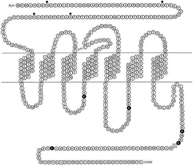
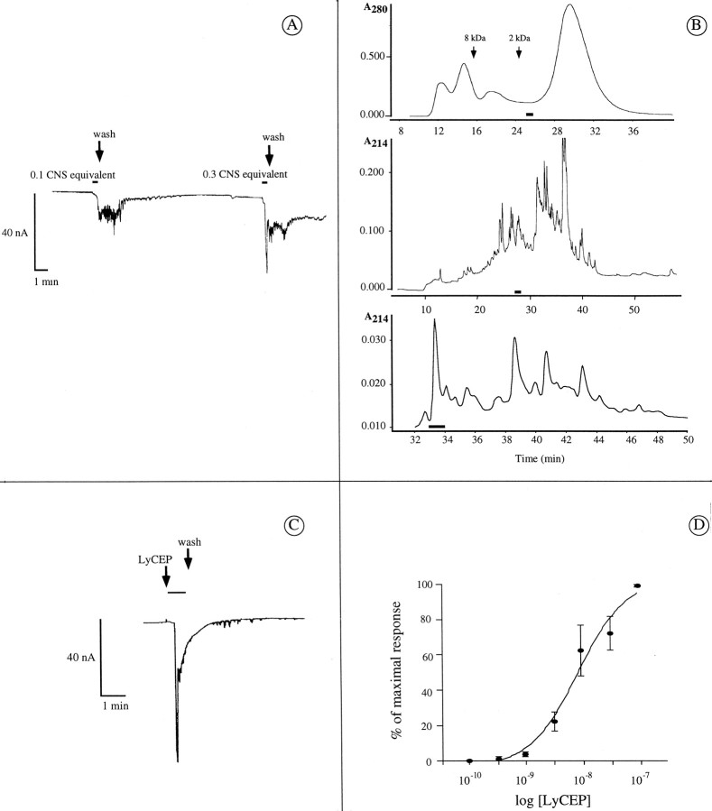
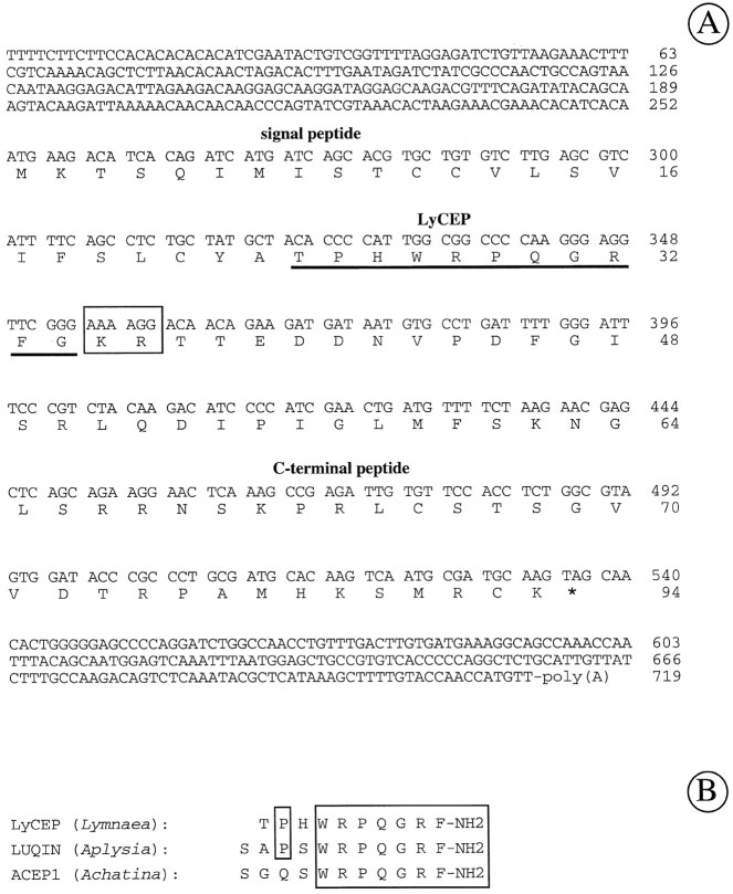
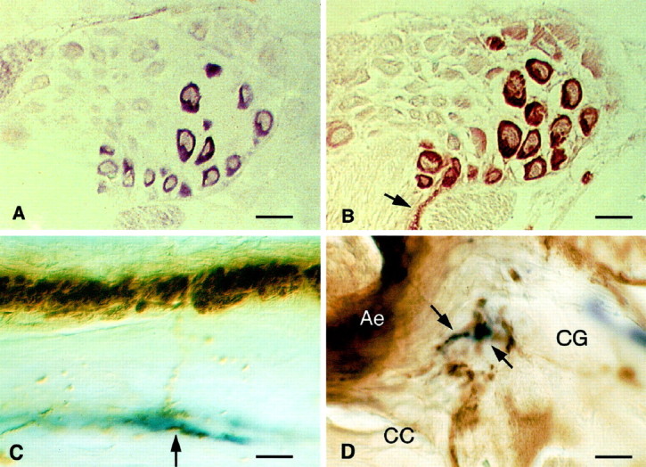
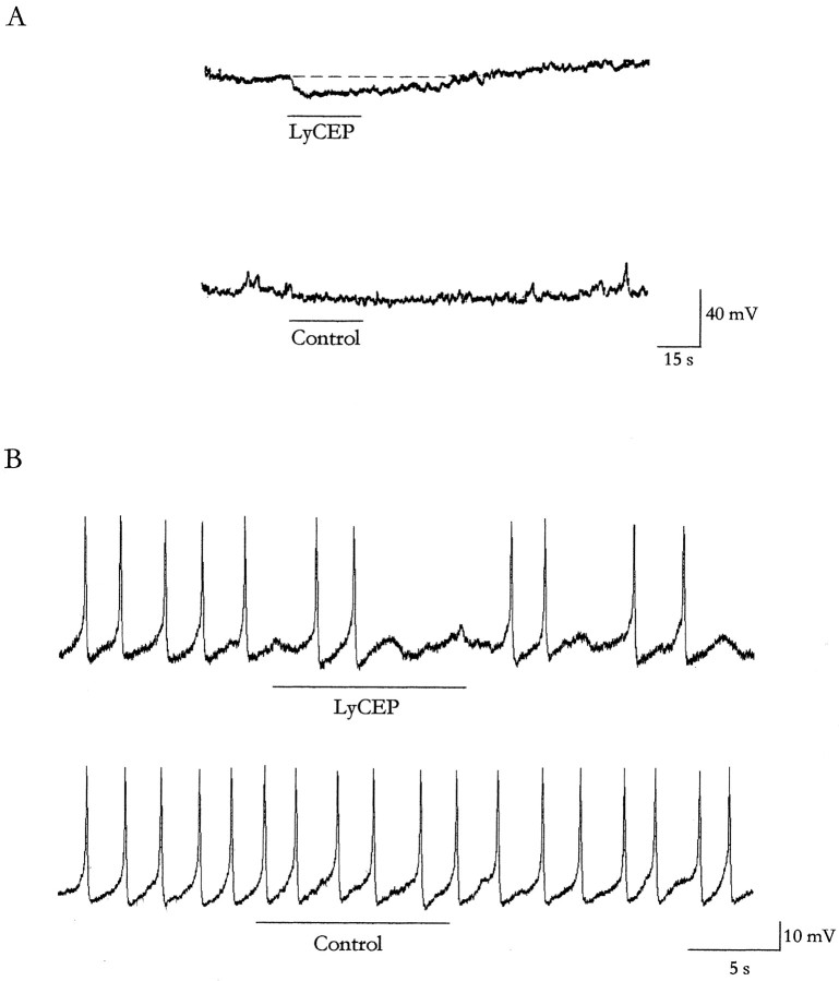
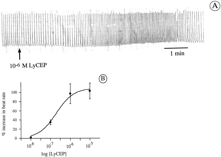
A novel G-protein-coupled receptor (GRL106) resembling neuropeptide Y and tachykinin receptors was cloned from the mollusc Lymnaea stagnalis. Application of a peptide extract from the Lymnaea brain to Xenopus oocytes expressing GRL106 activated a calcium-dependent chloride channel. Using this response as a bioassay, we purified the ligand for GRL106, Lymnaea cardioexcitatory peptide (LyCEP), an RFamide-type decapeptide (TPHWRPQGRF-NH2) displaying significant similarity to the Achatina cardioexcitatory peptide (ACEP-1) as well as to the recently identified family of mammalian prolactin-releasing peptides. In the Lymnaea brain, the cells that produce egg-laying hormone are the predominant site of GRL106 gene expression and appear to be innervated by LyCEP-containing fibers. Indeed, LyCEP application transiently hyperpolarizes isolated egg-laying hormone cells. In the Lymnaea pericardium, LyCEP-containing fibers end blindly at the pericardial lumen, and the heart is stimulated by LyCEP in vitro. These data confirm that LyCEP is an RFamide ligand for GRL106.


Figures

Fig. 1.
Fig. 1.
Deduced amino acid sequence and protein model of GRL106. Amino acids are given in single letter code. Potential N-linked glycosylation sites (dots) and putative protein kinase C (filled circles), casein kinase 2 (squares), and cAMP-dependent protein kinase phosphorylation (diamond) sites are indicated. The nucleotide sequence for GRL106 has been deposited into GenBank and is available under accession number AF037444.
Fig. 2.
Fig. 2.
Functional expression of GRL106 inXenopus oocytes and purification of the GRL106 ligand from Lymnaea brain extracts. A, Whole-cell current of a Xenopus oocyte expressing GRL106 and responding to a Lymnaea brain peptide extract. GRL106-encoding cRNA was injected into Xenopus oocytes, and after 48 hr whole-cell currents were measured while challenging the oocytes with a crude peptide extract from the LymnaeaCNS. Different amounts of the extract were applied for 25 sec (indicated by horizontal bars) and then washed out. One CNS equivalent is equal to an amount of extract that originates from one CNS. B, Purification of the bioactive peptide(s) from the brain extract. Top, HPGPC fractionation on Protein-Pak columns I-125 and I-300 connected in series (elution times of size markers are indicated).Middle, rpHPLC fractionation of the combined bioactive fractions of the HPGPC column on a Nucleosil C18 column eluted with 7.5 mm TFA and 0–60% acetonitrile. Bottom, rpHPLC fractionation of the combined bioactive fractions of the first rpHPLC column on a Nucleosil C18 column eluted with 0.05% HCl and 0–25% acetonitrile. Horizontal bars indicate bioactive fractions. C, Whole-cell current of aXenopus oocyte expressing GRL106 and responding to 3 nm synthetic LyCEP. D, Dose–response curve of the effect of synthetic LyCEP on whole-cell currents inXenopus oocytes expressing GRL106. LyCEP was applied in different concentrations to oocytes, and the mean membrane current of four experiments was plotted versus the logarithm of the concentration. Error bars denote SEM.
Fig. 3.
Fig. 3.
Nucleotide sequence of preproLyCEP cDNA, the derived amino acid sequence, and comparison of LyCEP with related peptides. A, Nucleotide sequence and conceptual translation of the LyCEP precursor cDNA. The putative proteolytic processing site (Lys-Arg) is boxed, and the LyCEP domain is underlined. Nucleotide numberis indicated at the right of each line. The predicted amino acid sequence of preproLyCEP isnumbered (on the right) with the first methionine designated position 1. The stop codon is indicated by anasterisk. The nucleotide sequence of the preproLyCEP cDNA has been deposited into GenBank under accession numberAF047683. B, Alignment of LyCEP with ACEP-1 fromAchatina fulica (Fujimoto et al., 1990) and with LUQIN from Aplysia californica (Aloyz and DesGroseillers, 1995). Amino acids identical in LyCEP and either of the other peptides are boxed.
Fig. 4.
Fig. 4.
Localization of GRL106 and LyCEP in theLymnaea CNS. A, GRL106 expression revealed by in situ hybridization of a section of theLymnaea CNS with a GRL106-specific probe. Within the cerebral ganglia, one of which is shown here, cells located at the position of CDC neurons hybridize with the GRL106 probe.B, Identification of CDCs by immunohistochemical staining of a section consecutive to the one in A with an antibody against ELH. The cells hybridizing with the GRL106 probe inA are clearly identified as CDCs by their strong reaction with the ELH antibody; in the commissure, immunopositive axon bundles and endings of the CDCs can be seen (arrow). Scale bars: A, B, 80 μm.C, Immunohistochemical double staining of theLymnaea CNS with an anti-ACEP-1 antibody (blue reaction product) and an anti-ELH antibody (brown reaction product). Shown is part of the cerebral commissure, where ELH-positive fibers seem to run to and make contact with fibers that react with the ACEP-1 antibody (arrow). Scale bar, 10 μm. D, In the neuropil of the cerebral ganglion (CG), axons that are immunoreactive to the ACEP-1 antibody and closely opposing axons containing ELH (arrows), which may indicate a site of contact between the LyCEP-expressing neurons and the CDCs. CC, Cerebral commissure containing the axon endings of the CDCs (Ae). Scale bar, 5 μm.
Fig. 5.
Fig. 5.
Effect of LyCEP on isolated CDC neurons.A, Effect of LyCEP on membrane potential. A representative intracellular recording of an isolated CDC at the resting membrane potential (−60 mV) to which 10 μm LyCEP (top) or vehicle (bottom) was applied under continuous superfusion. The horizontal lineindicates the moment and duration of application. B, Effect of LyCEP on spiking activity. A representative intracellular recording of an isolated CDC with spontaneous beating activity to which 5 μm LyCEP (top) or vehicle (bottom) was applied under continuous superfusion. Thehorizontal line indicates the moment and duration of application.
Fig. 6.
Fig. 6.
LyCEP immunoreactivity in the pericard. A strongly immunoreactive axon bundle (arrows) runs through the pericard (P). Inset, Occasionally small branches arise from this tract (asterisk, out of focus in this micrograph), penetrate the layer of mesothelial cells (Me; arrowhead), and project toward the pericardial cavity (Pc), which may suggest that the immunoreactive material is released into this cavity. A, Atrium. Scale bar: 100 μm; inset, 5 μm.
Fig. 7.
Fig. 7.
Effect of LyCEP on an isolatedLymnaea auricle. A, Contractions of an auricle that was dissected from the heart and placed in a displacement chamber. At the indicated time (arrow), 1 μm LyCEP was applied directly onto the auricle under continuous superfusion. B, Dose–response curve of the increase in beat rate induced by synthetic LyCEP on the isolated auricle. Each data point is the mean of at least seven determinations. Error bars denote SEM.

References

    1. Aloyz RS, DesGroseillers L. Processing of the L5–67 precursor peptide and characterization of LUQIN in the LUQ neurons of Aplysia californica. Peptides. 1995;16:331–338. - PubMed
    1. Bloem LJ, Yu L. A time-saving method for screening cDNA or genomic libraries. Nucleic Acids Res. 1990;18:2830. - PMC - PubMed
    1. Bradbury AF, Smyth DG. Peptide amidation. Trends Biochem Sci. 1991;16:112–115. - PubMed
    1. Brezina V, Eckert R, Erxleben C. Modulation of potassium conductances by an endogenous neuropeptide in neurones of Aplysia californica. J Physiol (Lond) 1987;382:267–290. - PMC - PubMed
    1. Brussaard AB, Kits KS, Ter Maat A, Van Minnen J, Moed PJ. Dual inhibitory action of FMRFamide on neurosecretory cells controlling egg laying behavior in the pond snail. Brain Res. 1988;447:35–51. - PubMed

Publication types

MeSH terms

LinkOut - more resources