. 1998 Dec 1;18(23):9896-909.
doi: 10.1523/JNEUROSCI.18-23-09896.1998.

Morphology of single geniculocortical afferents and functional recovery of the visual cortex after reverse monocular deprivation in the kitten

Affiliations

Morphology of single geniculocortical afferents and functional recovery of the visual cortex after reverse monocular deprivation in the kitten

A Antonini et al. J Neurosci. .

Abstract

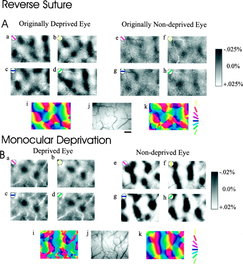
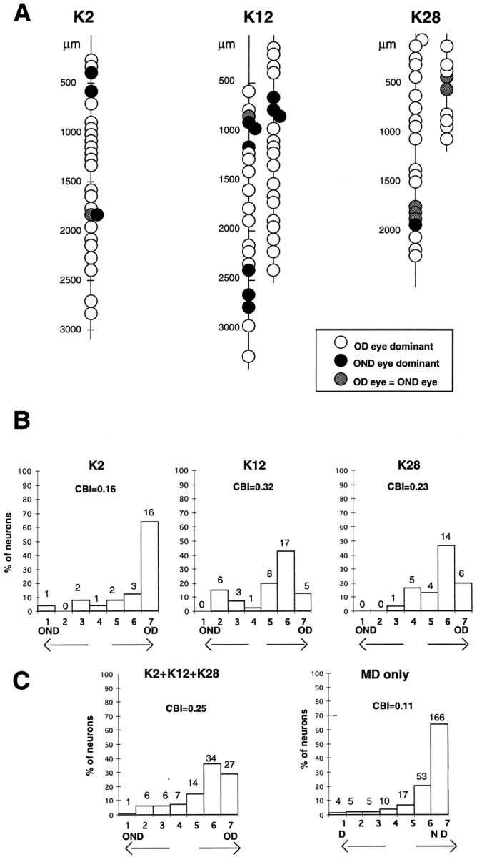
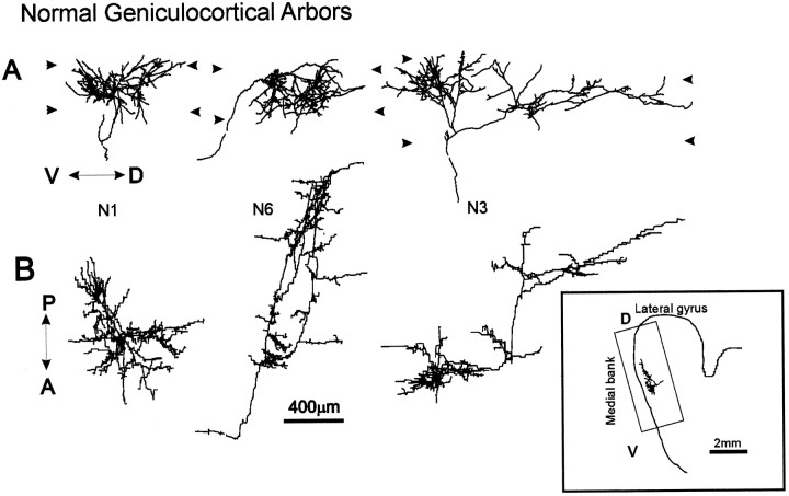
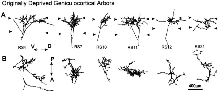
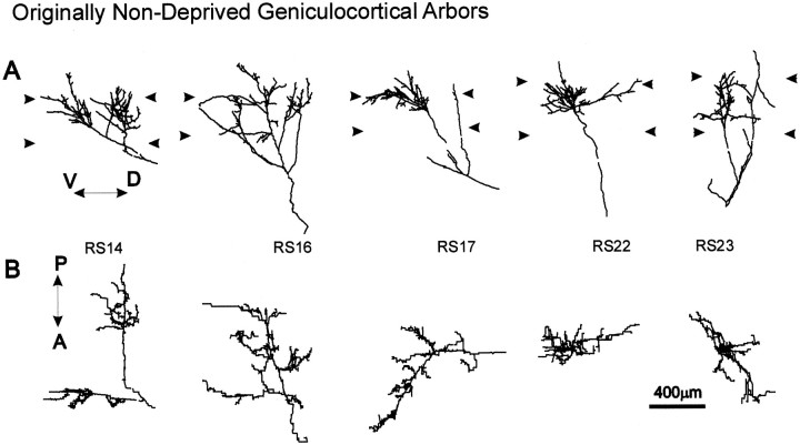
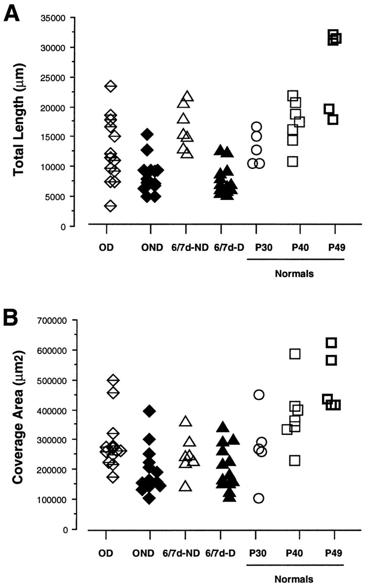
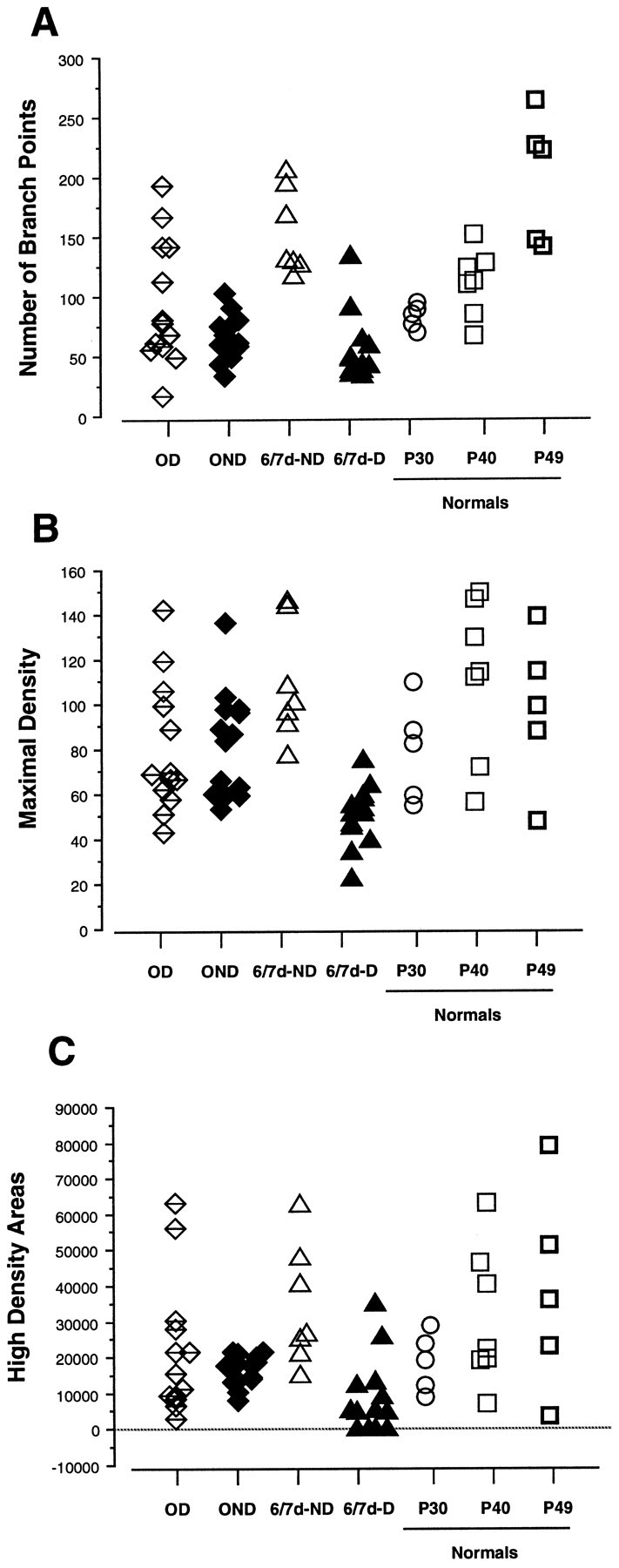
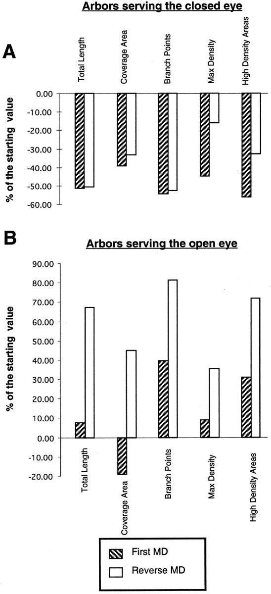
To investigate the possible anatomical basis for the functional recovery of visual cortical responses after reverse monocular deprivation, we have studied the morphology of single geniculocortical afferents to area 17. In kittens reverse-sutured for 10 d after an initial week of monocular deprivation, single-unit and intrinsic signal optical recordings confirmed that the effects of the initial deprivation were largely reversed. Responses through the originally nondeprived (OND) eye were drastically diminished, but remained much more selective for orientation than after an initial monocular deprivation (Crair et al., 1997). Responses through the originally deprived (OD) eye recovered completely. Geniculocortical afferent arbors in layer IV of area 17 were filled by iontophoresis of Phaseolus lectin into lamina A of the lateral geniculate nucleus (LGN) and were serially reconstructed. Arbors serving both the OD and the OND eye were analyzed. The plastic changes of both OD and OND arbors were evaluated by comparison with arbors reconstructed in normal animals and in animals studied after an equivalent initial period of deprivation (Antonini and Stryker, 1996). These analyses demonstrate that closure of the OND eye caused a substantial shrinkage of the arbors serving that eye. Moreover, reopening the OD eye induced regrowth only in some arbors, whereas others appeared to be largely unaffected and continued to have the characteristics of deprived arbors. Quantitatively, the initial and the second deprivation caused similar proportional changes in total arbor length and numbers of branches, whereas several other features were more severely affected by the initial deprivation.


Figures

Fig. 1.
Fig. 1.
Optical imaging of intrinsic signals obtained from the lateral gyrus in a reverse-suture kitten (A) and in a P38 kitten monocularly deprived for 7 d (B). In both A andB, a–h are cortical activity maps in response to each of the four oriented stimuli indicated in theleft corner of each image; j is the image of the vascular pattern, and i and k are color-coded angle maps of each hemisphere, combining the responses obtained from all orientations (see Materials and Methods).A, Distribution of cortical activity presented to the OD (a–d) and the OND eye (e–h). Note that the activity maps from the OND eye are orientation selective. i, k, Angle maps obtained from OD and OND eye, respectively.B, Distribution of cortical activity in response to oriented stimuli presented to the deprived eye (a–d) and the nondeprived eye (e–h). i, k, Angle maps obtained from deprived and nondeprived eye, respectively. Note in both the activity and angle maps that the responses through the deprived eye are nonoriented and limited to small cortical regions. Scale bar (shown in Aj for Aa–k, Ba–k): 500 μm.
Fig. 2.
Fig. 2.
Single units recorded in vertical penetrations along the medial bank of the lateral gyrus in three reverse-sutured animals. A, Sketch of electrode tracks with the single units sequentially encountered is shown for each animal. The dominance of the OD or OND eye in activating each cell is indicated by differentshading. Most neurons were dominated by the OD eye; units dominated by the OND eye, or equally driven by both eyes, were encountered in clusters. B, Ocular dominance distribution for neurons recorded in each animal. C, Ocular dominance distribution for the entire sample of cells recorded in the three reverse-sutured animals (left) and for 260 cells recorded in seven MD kittens [from Reiter and Stryker (1988)]. The contralateral bias index (CBI) is indicated above each plot.
Fig. 3.
Fig. 3.
Computer reconstructions of PHA-L-immunostained axonal arbors in area 17 in a normal P49 animal. All geniculocortical arbors in normal, OND (Fig. 4), and OD (Fig. 5) animals were obtained from the dorsal-most portion of the medial bank of the lateral gyrus.A shows the arbors as originally reconstructed in the coronal plane, and B shows the arbors as seen from the pial surface, after a 90° rotation along the dorsoventral axis of the lateral gyrus. The arrowheads indicate the boundaries of layer IV. V↔D = ventrodorsal axis indicated inA; A↔P = anteroposterior axis indicated in B. Inset, Drawing of coronal section showing arbor N3 and rectangle in the medial bank of the lateral gyrus containing all arbors reconstructed.Inset: D, Dorsal direction;V, ventral direction.
Fig. 4.
Fig. 4.
Computer reconstructions of PHA-L-immunostained geniculocortical arbors reconstructed in animals monocularly deprived for 1 week and reverse-sutured for 10 d. These arbors serve the OD eye. Arbors are shown in coronal view (A) and in surface view (B). Abbreviations and symbols are the same as in Figure 3.
Fig. 5.
Fig. 5.
Computer reconstructions of PHA-L-immunostained geniculocortical arbors serving the OND eye reconstructed in reverse-sutured animals. Arbors are shown in coronal view (A) and in surface view (B). Abbreviations and symbols are the same as in Figure 3.
Fig. 6.
Fig. 6.
Scattergram of the total length (A) and coverage area (B) of the terminal arborization in layer IV (see Materials and Methods) for arbors reconstructed in reverse-sutured animals (OD and OND arbors) and in the P49 normal control. For comparison, data from arbors serving the deprived and nondeprived eye in animals monocularly deprived for 6–7 d (6/7d-D and 6/7d-ND arbors) and in normal animals at P30 and P40 are also plotted.
Fig. 7.
Fig. 7.
Scattergrams of number of branch points (A), maximal-density (B), and (C) high-density areas (see Materials and Methods) of the terminal arborization in layer IV in arbors reconstructed in reverse-sutured animals (OD and OND arbors), in animals monocularly deprived for 6–7 d (6/7d-D and6/7d-ND arbors), and in normal animals at P30, P40, and P49. The density is expressed in μm/1000 μm2. Note that the high-density areas in four 6/7d-D arbors are equal to 0, because these arbors did not reach the threshold density of 38 μm/1000 μm2.
Fig. 8.
Fig. 8.
Percentage changes in total length, coverage area, number of branch points, maximal-density, and high-density areas after an initial period of 6–7 d of monocular deprivation (stipple bars) and after 10 d of reverse suture (white bars) in arbors serving the closed (A) and the open (B) eye. The percentage changes are referred to the state of the arbors before each deprivation. For the initial deprivation, comparisons were obtained relative to the values obtained in normal animals at P33 [(P33–6/7d-D) × 100/P33 and (P33–6/7d- ND) × 100/P33, for arbors serving the closed and open eye, respectively (see Results)]. For the reverse deprivation, we have measured the changes of OD and OND arbors relative to arbors reconstructed in animals deprived for 6–7 d [(6/7dND-OND × 100/6/7d-ND and (6/7d-D-OD) × 100/6/7d-D for arbors serving the closed and open eye, respectively]. Note that reverse suture has a greater effect than the initial deprivation on arbors serving the open eye, whereas arbors serving the closed eye appear to be equally affected by both the initial and reverse deprivation.

References

    1. Antonini A, Stryker MP. Rapid remodeling of axonal arbors in the visual cortex. Science. 1993a;260:1819–1821. - PubMed
    1. Antonini A, Stryker MP. Development of individual geniculocortical arbors in cat striate cortex and effects of binocular impulse blockade. J Neurosci. 1993b;13:3549–3573. - PMC - PubMed
    1. Antonini A, Stryker MP. Plasticity of geniculocortical afferents following brief or prolonged monocular occlusion in the cat. J Comp Neurol. 1996;369:64–82. - PubMed
    1. Antonini A, Stryker MP. Effect of sensory disuse on geniculate afferents to cat visual cortex. Vis Neurosci. 1998;15:401–409. - PMC - PubMed
    1. Blakemore C, Hawken MJ. Rapid restoration of functional input to the visual cortex of the cat after brief monocular deprivation. J Physiol (Lond) 1982;327:463–487. - PMC - PubMed

Publication types

Substances

LinkOut - more resources