. 1998 Dec 1;18(23):9996-10015.
doi: 10.1523/JNEUROSCI.18-23-09996.1998.

Neurons containing hypocretin (orexin) project to multiple neuronal systems

Affiliations

Neurons containing hypocretin (orexin) project to multiple neuronal systems

C Peyron et al. J Neurosci. .

Abstract

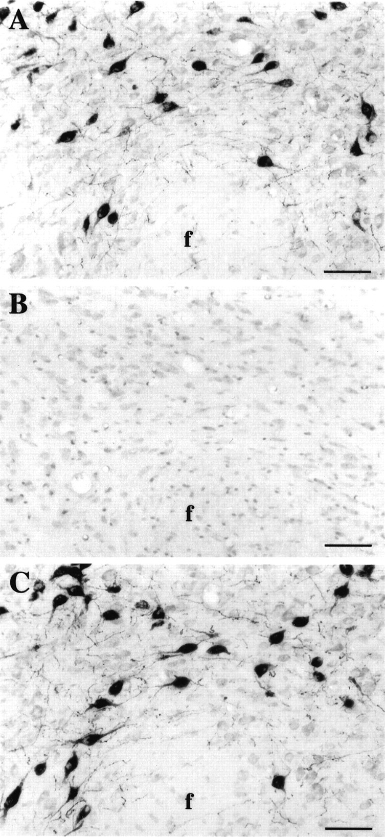
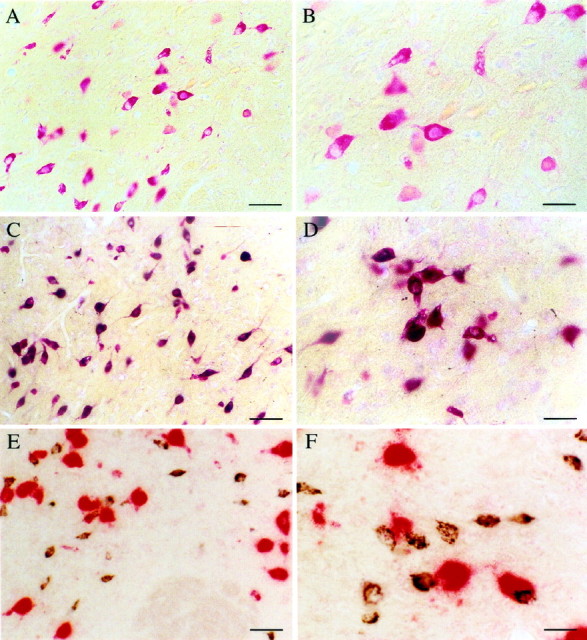
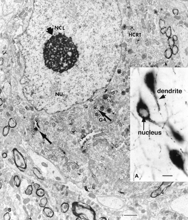
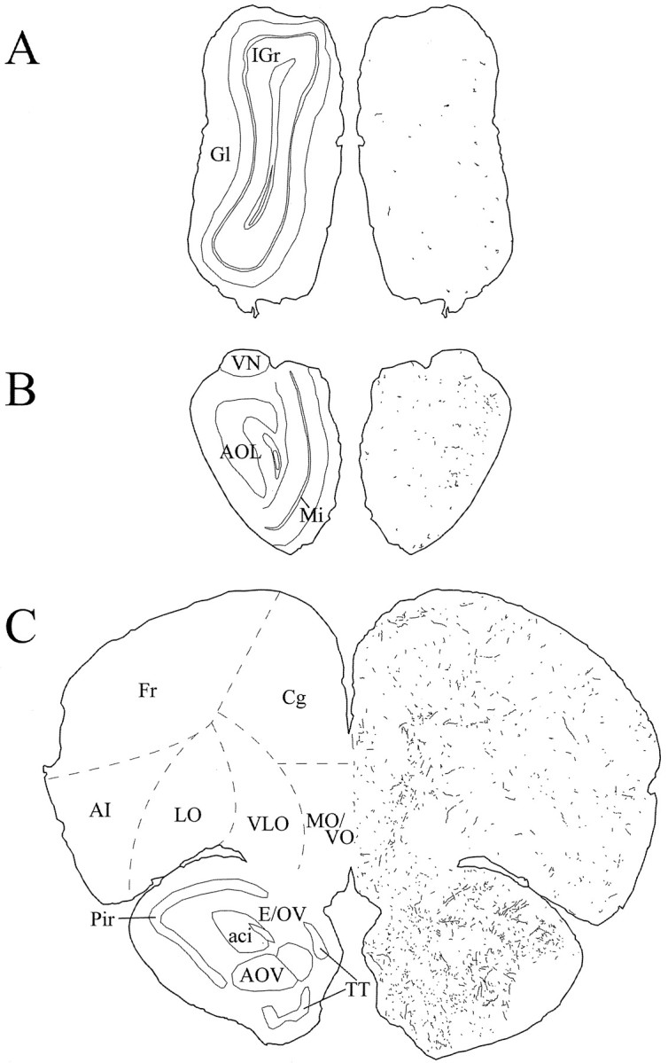
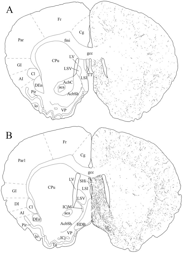
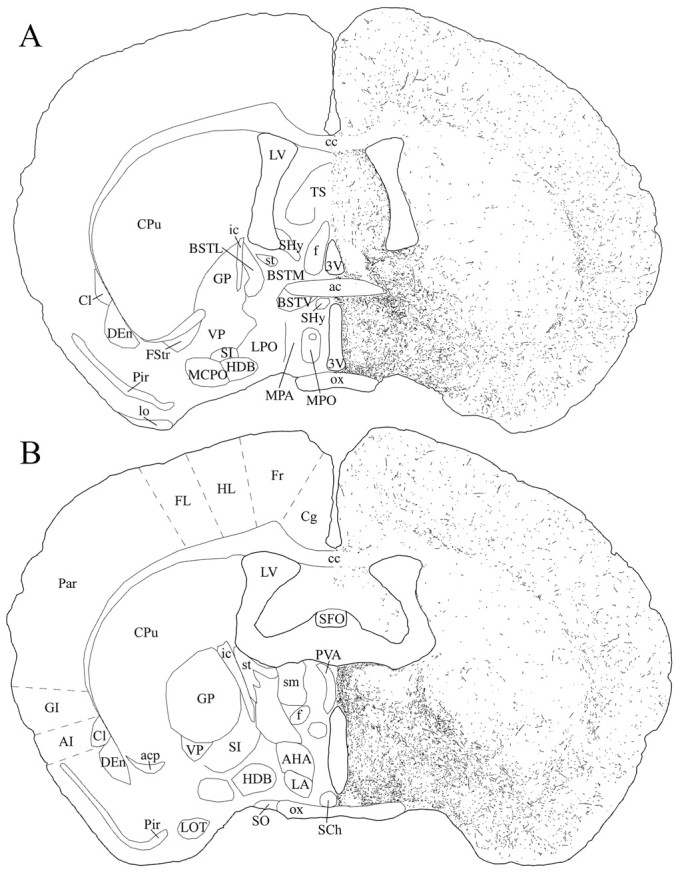
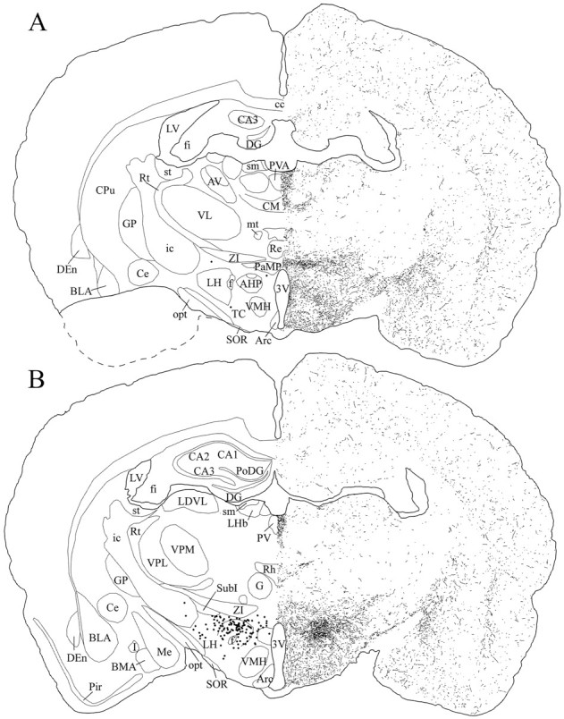
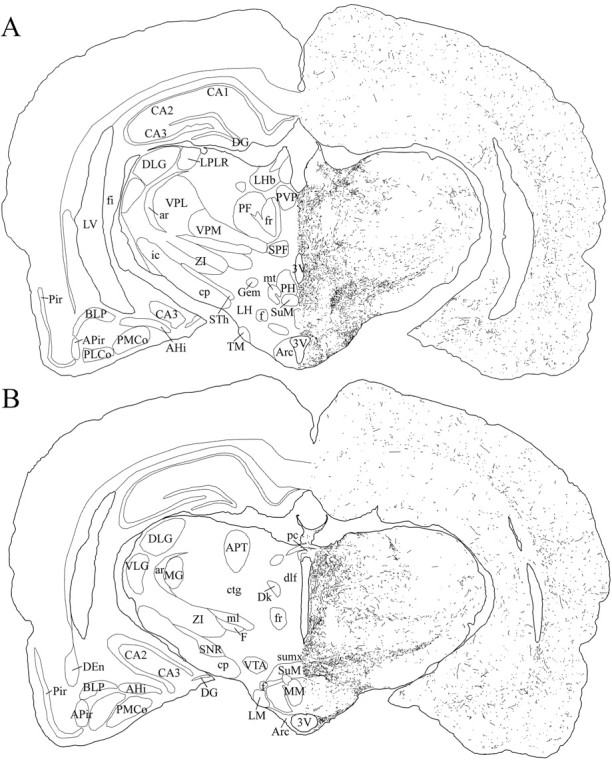
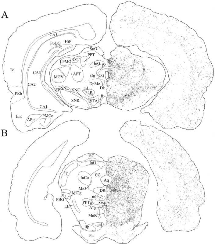
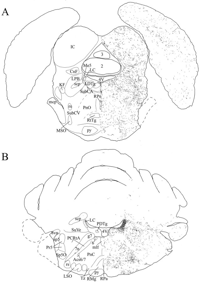
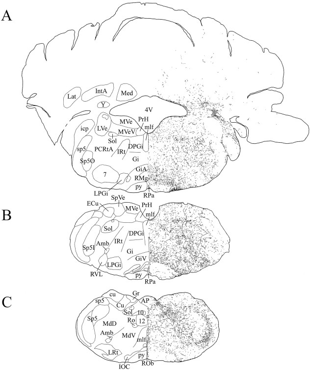
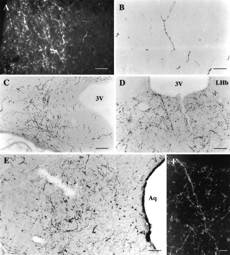
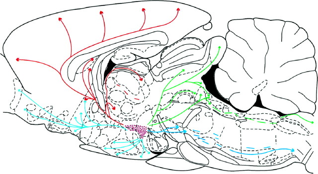
The novel neuropeptides called hypocretins (orexins) have recently been identified as being localized exclusively in cell bodies in a subregion of the tuberal part of the hypothalamus. The structure of the hypocretins, their accumulation in vesicles of axon terminals, and their excitatory effect on cultured hypothalamic neurons suggest that the hypocretins function in intercellular communication. To characterize these peptides further and to help understand what physiological functions they may serve, we undertook an immunohistochemical study to examine the distribution of preprohypocretin-immunoreactive neurons and fibers in the rat brain. Preprohypocretin-positive neurons were found in the perifornical nucleus and in the dorsal and lateral hypothalamic areas. These cells were distinct from those that express melanin-concentrating hormone. Although they represent a restricted group of cells, their projections were widely distributed in the brain. We observed labeled fibers throughout the hypothalamus. The densest extrahypothalamic projection was found in the locus coeruleus. Fibers were also seen in the septal nuclei, the bed nucleus of the stria terminalis, the paraventricular and reuniens nuclei of the thalamus, the zona incerta, the subthalamic nucleus, the central gray, the substantia nigra, the raphe nuclei, the parabrachial area, the medullary reticular formation, and the nucleus of the solitary tract. Less prominent projections were found in cortical regions, central and anterior amygdaloid nuclei, and the olfactory bulb. These results suggest that hypocretins are likely to have a role in physiological functions in addition to food intake such as regulation of blood pressure, the neuroendocrine system, body temperature, and the sleep-waking cycle.


Figures

Fig. 1.
Fig. 1.
Photomicrographs of adjacent frontal sections counterstained with neutral red at the level of the perifornical nucleus of the hypothalamus. A, Hcrt neurons were labeled using antiserum #2050 against the C-terminal 17 aa portion of the preproprotein. B, Hypocretin immunoreactivity was absent after preincubation of antiserum #2050 with the whole preproprotein, showing that this antibody specifically recognized hcrt.C, Hcrt neurons were labeled with antibody #2123. Identical staining was obtained with both antisera. f, Fornix. Scale bars, 65 μm.
Fig. 2.
Fig. 2.
Photomicrographs illustrating hcrt neurons in the perifornical nucleus of the hypothalamus. A, Neurons containing mRNA for hcrt visualized with a homogeneousred coloration of the cytoplasm with an in situ hybridization technique using Fast red as a substrate for alkaline phosphatase. B, Enlargement ofA. C, Photomicrograph showing that all neurons that stained red after in situhybridization (recognizing hcrt mRNA using Fast red) are labeledblack by immunohistochemistry (recognizing the protein with the antiserum #2050) for hcrt. This result indicates that the antiserum #2050 is specific for hcrt. D, High magnification of double-labeled cells after in situhybridization (red) and immunohistochemistry (black). E, Photomicrograph of neurons containing mRNA for the melanin-concentrating hormone (labeled inred by in situ hybridization using Vector red as substrate) and of hcrt neurons (labeled in blackby immunohistochemistry using DAB with nickel as the substrate). Note that no double-labeled cells are present, indicating that MCH and hcrt are found in two distinct populations of neurons. F, High magnification of the MCH (red) and hcrt (black) neurons in the perifornical nucleus. Scale bars:A, C, E, 65 μm;B, D, F, 36 μm.
Fig. 3.
Fig. 3.
Distribution of hcrt-labeled neurons on frontal sections at three rostrocaudal levels of the tuberal region of the hypothalamus (A to B to C) determined using #2123 antiserum. Sections were counterstained with neutral red (pink staining of all cells). The neutral red staining allowed us to determine the exact location of hcrt neurons in the brain that are localized exclusively in the tuberal region of the hypothalamus ventral to the zona incerta and that extend 1 mm rostrocaudally, beginning caudal to the paraventricular nucleus of the hypothalamus. 3V, 3rd ventricle; Arc, arcuate nucleus; DMH, DM, dorsomedial hypothalamic nucleus; f, fornix; ic, internal capsule; opt, optic tract; SOR, retrochiasmatic part of the supraoptic nucleus; VMH, ventromedial hypothalamic nucleus; ZI, zona incerta. Scale bars, 275 μm.
Fig. 4.
Fig. 4.
A, Large cells in the lateral hypothalamus are immunoreactive for hypocretin. Staining was found in the cytoplasm and dendrites (short arrow) but not in the nucleus (long arrow). Scale bar, 15 μm.B, Electron microscopic examination of immunoreactive neurons showed punctate staining in the cytoplasm, often associated with dense core granules and parts of the Golgi apparatus (thin arrow). Random organelles near dense core granules sometimes showed peroxidase label, probably because of diffusion during the process of immunocytochemistry. GA, Golgi apparatus; HCRT, hypocretin; NCL, nucleole (thick arrow); NU, nucleus.
Fig. 5.
Fig. 5.
Schematic drawings of 20 μm rostrocaudal coronal sections illustrating the distribution and relative density of hcrt fibers in the prefrontal cortex and the olfactory bulb after immunohistochemistry for hcrt using antibody #2050. aci, Anterior commissure, intrabulbar part;AI, agranular insular cortex; AOL, anterior olfactory nucleus, lateral part; AOV, anterior olfactory nucleus, ventral part; Cg, cingulate cortex;E/OV, ependyma and subependymal layer/olfactory ventricle; Fr, frontal cortex; Gl, glomerular layer of the olfactory bulb; IGr, internal granular layer of the olfactory bulb; LO, lateral orbital cortex; Mi, mitral cell layer of the olfactory bulb; MO/VO, medial/ventral orbital cortex;Pir, piriform cortex; TT, tenia tecta;VLO, ventrolateral orbital cortex; VN, vomeronasal nerve layer.
Fig. 6.
Fig. 6.
Schematic drawings of 20 μm rostrocaudal coronal sections illustrating the distribution and relative density of hcrt fibers in the telencephalon after immunohistochemistry for hcrt using antibody #2050. aca, Anterior commissure, anterior part;AcbC, accumbens nucleus, core; AcbSh, accumbens nucleus, shell; AI, agranular insular cortex;Cg, cingulate cortex; Cl, claustrum;CPu, caudate putamen; DEn, dorsal endopiriform nucleus; DI, dysgranular insular cortex;fmi, forceps minor of the corpus callosum;Fr, frontal cortex; gcc, genu of the corpus callosum; GI, granular insular cortex;HDB, nucleus of the horizontal limb of the diagonal band; ICj, islands of Calleja; ICjM, islands of Calleja, major island; lo, lateral olfactory tract; LSI, lateral septal nucleus, intermediate part;LSV, lateral septal nucleus, ventral part;LV, lateral ventricle; Par, parietal cortex; Par1, parietal cortex, area 1;Pir, piriform cortex; SHi, septohippocampal nucleus; TT, tenia tecta;Tu, olfactory tubercle; VP, ventral pallidum.
Fig. 7.
Fig. 7.
Schematic drawings of 20 μm rostrocaudal coronal sections illustrating the distribution and relative density of hcrt fibers at the level of the preoptic area after immunohistochemistry for hcrt using antibody #2050. 3V, 3rd ventricle;ac, anterior commissure; acp, anterior commissure, posterior part; AHA, anterior hypothalamic area, anterior part; AI, agranular insular cortex;BSTL, bed nucleus of the stria terminalis, lateral division; BSTM, bed nucleus of the stria terminalis, medial division; BSTV, bed nucleus of the stria terminalis, ventral division; cc, corpus callosum;Cg, cingulate cortex; Cl, claustrum; CPu, caudate putamen; DEn, dorsal endopiriform nucleus;f, fornix; FL, forelimb area of the cortex; Fr, frontal cortex; FStr, fundus striati; GI, granular insular cortex; GP, globus pallidus; HDB, nucleus of the horizontal limb of the diagonal band; HL, hindlimb area of the cortex;ic, internal capsule; LA, lateroanterior hypothalamic nucleus; lo, lateral olfactory tract;LOT, nucleus of the lateral olfactory tract;LPO, lateral preoptic area; LV, lateral ventricle; MCPO, magnocellular preoptic nucleus;MPA, medial preoptic area; MPO, medial preoptic nucleus; ox, optic chiasm; Par, parietal cortex; Pir, piriform cortex;PVA, paraventricular thalamic nucleus, anterior part;SCh, suprachiasmatic nucleus; SFO, subfornical organ; SHy, septohypothalamic nucleus;SI, substantia innominata; sm, stria medullaris of the thalamus; SO, supraoptic nucleus;st, stria terminalis; TS, triangular septal nucleus; VP, ventral pallidum.
Fig. 8.
Fig. 8.
Schematic drawings of 20 μm rostrocaudal coronal sections illustrating the distribution and relative density of hcrt fibers at the level of the tuberal region of the hypothalamus after immunohistochemistry for hcrt using antibody #2050. The position of hcrt cell bodies is indicated as dots in the left hemisphere. 3V, 3rd ventricle; AHP, anterior hypothalamic area, posterior part; Arc, arcuate nucleus; AV, anteroventral thalamic nucleus;BLA, basolateral amygdaloid nucleus, anterior part;BMA, basomedial amygdaloid nucleus, anterior part;CA1–CA3, fields CA1–CA3 of Ammon’s horn;cc, corpus callosum; Ce, central amygdaloid nucleus; CM, central medial thalamic nucleus;CPu, caudate putamen; DEn, dorsal endopiriform nucleus; DG, dentate gyrus;f, fornix; fi, fimbria of the hippocampus; G, gelatinosus thalamic nucleus;GP, globus pallidus; I, intercalated nuclei of the amygdala; ic, internal capsule;LDVL, laterodorsal thalamic nucleus, ventrolateral part;LH, lateral hypothalamic area; LHb, lateral habenular nucleus; LV, lateral ventricle;Me, medial amygdaloid nucleus; mt, mammillothalamic tract; opt, optic tract;PaMP, paraventricular hypothalamic nucleus, medial parvocellular part; Pir, piriform cortex;PoDG, polymorph layer of the dentate gyrus;PV, paraventricular thalamic nucleus;PVA, paraventricular thalamic nucleus, anterior part;Re, reuniens thalamic nucleus; Rh, rhomboid thalamic nucleus; Rt, reticular thalamic nucleus; sm, stria medullaris of the thalamus;SOR, supraoptic nucleus, retrochiasmatic part;st, stria terminalis; SubI, subincertal nucleus; TC, tuber cinereum area; VL, ventrolateral thalamic nucleus; VMH, ventromedial hypothalamic nucleus; VPL, ventral posterolateral thalamic nucleus; VPM, ventral posteromedial thalamic nucleus; ZI, zona incerta.
Fig. 9.
Fig. 9.
Schematic drawings of 20 μm rostrocaudal coronal sections illustrating the distribution and relative density of hcrt fibers in the caudal part of the hypothalamus after immunohistochemistry for hcrt using antibody #2050. 3V, 3rd ventricle; AHi, amygdalohippocampal area;APir, amygdalopiriform transition area;APT, anterior pretectal nucleus; ar, acoustic stria; Arc, arcuate nucleus;BLP, basolateral amygdaloid nucleus, posterior part;CA1–CA3, fields CA1–CA3 of Ammon’s horn;cp, cerebral peduncle; ctg, central tegmental tract; DEn, dorsal endopiriform nucleus;DG, dentate gyrus; Dk, nucleus Darkschewitsch; dlf, dorsal longitudinal fasciculus; DLG, dorsal lateral geniculate nucleus;f, fornix; F, nucleus of the fields of Forel; fi, fimbria of the hippocampus;fr, fasciculus retroflexus; Gem, gemini hypothalamic nucleus; ic, internal capsule;LH, lateral hypothalamic area; LHb, lateral habenular nucleus; LM, lateral mammillary nucleus; LPLR, lateral posterior thalamic nucleus, laterorostral part; LV, lateral ventricle;MG, medial geniculate nucleus; ml, medial lemniscus; MM, medial mammillary nucleus, medial part;mt, mammillothalamic tract; pc, posterior commissure; PF, parafascicular thalamic nucleus;PH, posterior hypothalamic area; Pir, piriform cortex; PLCo, posterolateral cortical amygdaloid nucleus; PMCo, posteromedial cortical amygdaloid nucleus; PVP, paraventricular thalamic nucleus, posterior part; SNR, substantia nigra, reticular part; SPF, subparafascicular thalamic nucleus;STh, subthalamic nucleus; SuM, supramammillary nucleus; sumx, supramammillary decussation; TM, tuberomammillary nucleus;VLG, ventral lateral geniculate nucleus;VPL, ventral posterolateral thalamic nucleus;VPM, ventral posteromedial thalamic nucleus;VTA, ventral tegmental area; ZI, zona incerta.
Fig. 10.
Fig. 10.
Schematic drawings of 20 μm rostrocaudal coronal sections illustrating the distribution and relative density of hcrt fibers in the midbrain after immunohistochemistry for hcrt using antibody #2050. APir, Amygdalopiriform transition area;APT, anterior pretectal nucleus; Aq, aqueduct; ATg, anterior tegmental nucleus;CA1–CA3, fields CA1–CA3 of Ammon’s horn;CG, central gray; cp, cerebral peduncle;ctg, central tegmental tract; Dk, nucleus Darkschewitsch; DpMe, deep mesencephalic nucleus; DR, dorsal raphe nucleus; Ent, entorhinal cortex; fr, fasciculus retroflexus;HiF, hippocampal fissure; IC, inferior colliculus; InCo, intercollicular nucleus;InG, intermediate gray layer of the superior colliculus;lfp, longitudinal fasciculus of the pons;LL, lateral lemniscus; LPMC, lateral posterior thalamic nucleus, mediocaudal part; Me5, mesencephalic trigeminal nucleus; MGV, medial geniculate nucleus, ventral part; MiTg, microcellular tegmental nucleus; ml, medial lemniscus; mlf, medial longitudinal fasciculus; MnR, median raphe nucleus; OT, nucleus of the optic tract;PBG, parabigeminal nucleus; pc, posterior commissure; PMCo, posteromedial cortical amygdaloid nucleus; Pn, pontine nuclei; PoDG, polymorph layer of the dentate gyrus; PPT, posterior pretectal nucleus; PPTg, pedunculopontine tegmental nucleus; PRh, perirhinal cortex; R, red nucleus; SC, superior colliculus; SNC, substantia nigra, compact part; SNL, substantia nigra, lateral part; SNR, substantia nigra, reticular part;SuG, superficial gray layer of the superior colliculus;Te, temporal cortex; VTA, ventral tegmental area; xscp, decussation of the superior cerebellar peduncle.
Fig. 11.
Fig. 11.
Schematic drawings of 20 μm rostrocaudal coronal sections illustrating the distribution and relative density of hcrt fibers in the pons after immunohistochemistry for hcrt using antibody #2050. 2,3, Cerebellar lobules;4V, 4th ventricle; 6, abducens nucleus;7n, facial nerve or its root; 8vn, vestibular root, vestibulocochlear nerve; Acs6/7, accessory abducens and facial nuclei; CnF, cuneiform nucleus; g7, genu of the facial nerve;IC, inferior colliculus; KF, Kölliker–Fuse nucleus; LC, locus coeruleus;LDTg, laterodorsal tegmental nucleus;LPB, lateral parabrachial nucleus; LSO, lateral superior olive;mcp, middle cerebellar peduncle; Me5, mesencephalic trigeminal nucleus; mlf, medial longitudinal fasciculus; MSO, medial superior olive;PCRtA, parvocellular reticular nucleus, α part;PDTg, posterodorsal tegmental nucleus;PnC, pontine reticular nucleus, caudal part;PnO, pontine reticular nucleus, oral part;Pr5, principal sensory trigeminal nucleus;py, pyramidal tract; RMg, raphe magnus nucleus; RPa, raphe pallidus nucleus;RPn, raphe pontis nucleus; rs, rubrospinal tract; RtTg, reticulotegmental nucleus of the pons; scp, superior cerebellar peduncle;sp5, spinal trigeminal tract; Sp5O, spinal trigeminal nucleus, oral part; SubCA, subcoeruleus nucleus, α part; SubCV, subcoeruleus nucleus, ventral part; SuVe, superior vestibular nucleus; Tz, nucleus of the trapezoid body.
Fig. 12.
Fig. 12.
Schematic drawings of 20 μm rostrocaudal coronal sections illustrating the distribution and relative density of hcrt fibers in the medulla after immunohistochemistry for hcrt using antibody #2050. 4V, 4th ventricle; 7, facial nucleus; 10, dorsal motor nucleus of vagus;12, hypoglossal nucleus; Amb, ambiguus nucleus; AP, area postrema;cu, cuneate fasciculus; Cu, cuneate nucleus; DPGi, dorsal paragigantocellular nucleus;ECu, external cuneate nucleus; Gi, gigantocellular reticular nucleus; GiA, gigantocellular reticular nucleus, α part; GiV, gigantocellular reticular nucleus, ventral part; Gr, gracile nucleus;icp, inferior cerebellar peduncle; IntA, interposed cerebellar nucleus, anterior part; IOC, inferior olive, subnucleus C of medial nucleus; IRt, intermediate reticular nucleus; Lat, lateral cerebellar nucleus; LPGi, lateral paragigantocellular nucleus;LRt, lateral reticular nucleus; LVe, lateral vestibular nucleus; MdD, medullary reticular nucleus, dorsal part; MdV, medullary reticular nucleus, ventral part; Med, medial cerebellar nucleus;mlf, medial longitudinal fasciculus; MVe, medial vestibular nucleus; MVeV, medial vestibular nucleus, ventral part; PCRtA, parvocellular reticular nucleus, α part; PrH, prepositus hypoglossal nucleus;py, pyramidal tract; RMg, raphe magnus nucleus; Ro, nucleus of Roller; ROb, raphe obscurus nucleus; RPa, raphe pallidus nucleus;RVL, rostroventrolateral reticular nucleus;Sol, nucleus of the solitary tract; sp5, spinal trigeminal tract; Sp5, spinal trigeminal nucleus;Sp5I, spinal trigeminal nucleus, interpolar part;Sp5O, spinal trigeminal nucleus, oral part;SpVe, spinal vestibular nucleus; Y, nucleus Y.
Fig. 13.
Fig. 13.
Photomicrographs of hcrt immunoreactive axons in the rat brain using the antiserum #2123. A, Dark-field illustration of thick hcrt fibers (in white) located in the locus coeruleus, lateral to the 4th ventricle. Notice that fibers are restricted to the locus and contain numerous boutons.B, Photomicrograph showing that hcrt-IR fibers were mainly long with varicosities. The density of fibers was relatively low in all cortical areas as shown in this picture of the frontal cortex.C, Illustration of the numerous long and thick fibers seen in the caudal part of the arcuate nucleus. Hcrt fibers contain numerous boutons. D, F, Photomicrographs illustrating one of the main projections for hcrt neurons, the paraventricular nucleus of the thalamus. Fibers were long with numerous varicosities as illustrated in F on a dark-field enlargement of D. E, Photomicrograph showing the high density of varicose fibers located in the lateral periaqueductal gray at the level of the dorsal raphe nucleus. Scale bars: A, 36 μm; BD, 70 μm; E, 50 μm; F, 25 μm.3V, 3rd ventricle; Aq, Aqueduct;LHb, lateral habenular nucleus.
Fig. 14.
Fig. 14.
Schematic summary drawing of pathways taken by hcrt processes that widely innervate rat brain. The sagittal section used is taken from the atlas of Paxinos and Watson (1986).Purple dots: Hypocretin-labeled neurons;red: dorsal ascending pathway; light blue: ventral ascending pathway; green: dorsal descending pathway; dark blue: ventral descending pathway.

References

    1. Abercrombie M. Estimation of nuclear population from microtome sections. Anat Rec. 1946;94:239–247. - PubMed
    1. Akabayashi A, Wahlestedt C, Alexander JT, Leibowitz SF. Specific inhibition of endogenous neuropeptide Y synthesis in arcuate nucleus by antisense oligonucleotides suppresses feeding behavior and insulin secretion. Mol Brain Res. 1994;21:55–61. - PubMed
    1. Allen GV, Cechetto DF. Functional and anatomical organization of cardiovascular pressor and depressor sites in the lateral hypothalamic area: I. Descending projections. J Comp Neurol. 1992;315:313–332. - PubMed
    1. Allen GV, Cechetto DF. Functional and anatomical organization of cardiovascular pressor and depressor sites in the lateral hypothalamic area: II. Ascending projections. J Comp Neurol. 1993;330:421–438. - PubMed
    1. Bittencourt JC, Presse F, Arias C, Peto C, Vaughan J, Nahon JL, Vale W, Sawchenko PE. The melanin-concentrating hormone system of the rat brain: an immuno- and hybridization histochemical characterization. J Comp Neurol. 1992;319:218–245. - PubMed

Publication types

MeSH terms

LinkOut - more resources