. 1998 Dec 1;18(23):10136-49.
doi: 10.1523/JNEUROSCI.18-23-10136.1998.

Immunocytochemical localization of the postsynaptic density protein PSD-95 in the mammalian retina

Affiliations

Immunocytochemical localization of the postsynaptic density protein PSD-95 in the mammalian retina

P Koulen et al. J Neurosci. .

Abstract

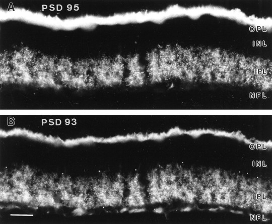
Synapse-associated proteins are the scaffold for the selective aggregation of ion channels at synapses; they provide the link to cytoskeletal elements and possibly are involved with the regulation of synaptic efficacy by electrical activity. The localization of the postsynaptic density protein PSD-95 was studied in different mammalian retinae (rat, monkey, and tree shrew) by using immunocytochemical methods. Immunofluorescence for PSD-95 was most prominent in the outer plexiform layer (OPL). The axon terminals of rods and cones, the rod spherules and cone pedicles, were strongly labeled. Electron microscopy, using preembedding immunocytochemistry, showed PSD-95 localized presynaptically within the photoreceptor terminals. Distinct PSD-95 labeling was also present in the inner plexiform layer (IPL). It had a punctate appearance suggesting the synaptic clustering of PSD-95 in the IPL. Electron microscopy showed that PSD-95 was concentrated in processes that were postsynaptic at bipolar cell ribbon synapses (dyads). As a rule, only one of the two postsynaptic members of the dyad was labeled for PSD-95. Double-labeling experiments were performed for PSD-95 and for SAP 102 or PSD-93, respectively, two other members of the family of synapse-associated proteins. All three were found to be colocalized in the synaptic hot spots in the IPL. In the OPL, however, PSD-95 and PSD-93 were found presynaptically, whereas SAP 102 was located postsynaptically at photoreceptor synapses. Double-labeling experiments also were performed for PSD-95 and for the NR1 subunit of the NMDA receptor. They were found to be colocalized in synaptic hot spots in the IPL.


Figures

Fig. 1.
Fig. 1.
Photomicrograph of a vertical cryostat section through a rat retina that was immunolabeled for PSD-95. The retinal layers are shown in the Nomarski micrograph (right).ONL, Outer nuclear layer; OPL, outer plexiform layer; INL, inner nuclear layer;IPL, inner plexiform layer; GCL, ganglion cell layer. PSD-95 immunofluorescence is prominent in the OPL, where the axon terminals of rods, the so-called rod spherules, are labeled intensively. In the IPL punctate immunofluorescence is found, representing a concentration of PSD-95 at synapses. Scale bar, 25 μm.
Fig. 2.
Fig. 2.
High-power fluorescence micrographs of vertical cryostat sections through rat (A), macaque monkey (B), and tree shrew (C) retinae that were immunostained for PSD-95. A, Rod spherules at the outer margin of the OPL are labeled in the rat.B, Four cone pedicles (arrows) and several rod spherules are labeled in the monkey retina.C, Exclusively, cone pedicles are labeled in the tree shrew retina. The labeling in rod spherules and cone pedicles is concentrated in their outer membranes.
Fig. 3.
Fig. 3.
Electron micrographs of sections through cone pedicles (A, C) and rod spherules (B, D) of a rat retina that was immunolabeled for PSD-95. A, The labeled cone pedicle base (top half) contains two synaptic ribbons (arrowheads) and receives several invaginating processes (stars) from unlabeled bipolar or horizontal cell dendrites (bottom half). B, Section of the synaptic triad within a labeled rod spherule. Thearrowhead indicates the ribbon; the three invaginating processes (stars) are unlabeled. C, The labeled cone pedicle base (top half) contains three synaptic ribbons and receives several unlabeled invaginating processes.D, Section of the synaptic triad within a labeled rod spherule. The arrowhead indicates the ribbon; thestars mark unlabeled invaginating processes. Scale bars, 0.3 μm.
Fig. 4.
Fig. 4.
Electron micrographs of sections through the IPL of a rat retina that was immunolabeled for PSD-95. A–C, Shown are the axon terminals of putative OFF cone bipolar cells from the outer part of the IPL. The presynaptic ribbons are marked byarrowheads. Only one of the two postsynaptic processes at this ribbon synapse (dyad) appears to be labeled (star). D–F, Shown are the axon terminals of putative ON cone bipolar cells from the inner part of the IPL. The presynaptic ribbons are marked by arrowheads. Labeling is found in only one of the two postsynaptic processes.G–I, Shown are the axon terminals of putative rod bipolar cells close to the ganglion cell layer. The presynaptic ribbons are opposed to two postsynaptic processes, of which one (star) appears to be labeled. Scale bars, 0.2 μm.
Fig. 5.
Fig. 5.
Vertical cryostat section through a rat retina double-labeled for PKC (A) and PSD-95 (B). The fluorescence micrographs show the inner one-third of the IPL. A, PKC-immunolabeled axon terminals of rod bipolar cells form small varicosities 2–3 μm in diameter. Three are indicated by arrows.B, PSD-95 immunofluorescence is concentrated in “hot spots” 0.25–1 μm in diameter. The hot spots are aggregated in groups (arrows) that correspond to the varicosities inA. Careful inspection of this high-power light micrograph shows that neighboring hot spots can still be resolved if their distance is ∼0.3 μm.
Fig. 6.
Fig. 6.
Vertical cryostat section through a rat retina double-labeled for PSD-95 (A) and SAP 102 (B). The Nomarski micrograph in Cshows the retinal layers. A, PSD-95 immunofluorescence is most prominent in rod spherules of the OPL. In the IPL the fluorescence is concentrated in “hot spots.” B, SAP 102 immunofluorescence is sparse in the OPL, and weak extrasynaptic labeling is present in the INL; in the IPL the labeling is concentrated in hot spots, and prominent labeling is also present in the optic nerve fiber layer (NFL). Because of the short fixation the tissue was very soft, and the cryostat sections were of uneven thickness and appeared to be ruptured.
Fig. 7.
Fig. 7.
High-power fluorescence micrograph of a vertical section through the OPL of a rat retina that was double-labeled for PSD-95 (A) and SAP 102 (B).A, Slightly blurred rod spherules express PSD-95 immunofluorescence. B, Small puncta express SAP 102 immunoreactivity. We previously have shown by electron microscopy that these puncta represent terminals of horizontal cell processes (Koulen et al., 1998). C, The same micrograph as inA, with the puncta, taken from B, inserted as black dots.
Fig. 8.
Fig. 8.
The high-power photomicrographs (left column, A, D, G) show the IPL of double-labeled sections through rat retinae. PSD-95 immunofluorescence is shown in theleft side of each micrograph. Immunofluorescence for SAP 102 (A), PSD-93 (D), and NR1 (G) are shown on the right side and are printed as mirror images. The midline is cut along a common border, and puncta at symmetric positions with respect to the midline represent colocalizations. Drawings of all puncta are shown in the middle column (B, E, H). Drawings of puncta at symmetrical positions, representing colocalizations, are shown in the right column(C, F, I). The numbers of the labeled puncta are inserted. Each frame represents 40 × 40 μm.
Fig. 9.
Fig. 9.
Vertical cryostat section through a rat retina double-labeled for PSD-95 (A) and PSD-93 (B). The labeling in the OPL is much more prominent for PSD-95; however, close inspection in the microscope shows that both PSD-95 and PSD-93 label rod spherules. Punctate labeling is present in the IPL both in A and B, and comparison of the puncta suggests that they are colocalized. This is demonstrated in more detail in Figure 8D. The optic nerve fiber layer expresses PSD-93 (labeling at the bottom ofB), but not PSD-95. Because of the short fixation the IPL appears to be ruptured. Scale bar, 25 μm.
Fig. 10.
Fig. 10.
Vertical cryostat section through a rat retina double-labeled for PSD-95 (A) and the NR1 subunit of the NMDA receptor (B). In addition to the punctate labeling in the IPL, NR1 immunofluorescence is also present in cell bodies of the ganglion cell layer and in some amacrine cells (arrow). Weak and sparse labeling is also present in the OPL; a putative horizontal cell body is indicated by thearrowhead. Because of the short fixation the IPL appears to be ruptured. Scale bar, 25 μm.

References

    1. Barnes S. After transduction: response shaping and control of transmission by ion channels of the photoreceptor inner segment. Neuroscience. 1994;58:447–459. - PubMed
    1. Brandstätter JH, Hartveit E, Sassoè-Pognetto M, Wässle H. Expression of NMDA and high-affinity kainate receptor subunit mRNAs in the adult rat retina. Eur J Neurosci. 1994;6:1100–1112. - PubMed
    1. Brandstätter JH, Koulen P, Kuhn R, van der Putten H, Wässle H. Compartmental localization of a metabotropic glutamate receptor (mGluR7): two different active sites at a retinal synapse. J Neurosci. 1996;16:4749–4756. - PMC - PubMed
    1. Brandstätter JH, Koulen P, Wässle H. Selective synaptic distribution of kainate receptor subunits in the two plexiform layers of the rat retina. J Neurosci. 1997;17:9298–9307. - PMC - PubMed
    1. Brandstätter JH, Koulen P, Wässle H. Diversity of glutamate receptors in the mammalian retina. Vision Res. 1998;38:1385–1397. - PubMed

Publication types

MeSH terms