. 1998 Oct;43(4):519-24.
doi: 10.1136/gut.43.4.519.

Mucin gene expression in human embryonic and fetal intestine

Affiliations

Mucin gene expression in human embryonic and fetal intestine

M P Buisine et al. Gut. 1998 Oct.

Abstract

Background: The intestinal epithelium is covered by a continuous layer of mucus which is secreted by well differentiated epithelial cells. Disregulation of the expression of mucins has been reported to have possible implications in the neoplastic process which affects intestinal mucosae. It is well known that preneoplastic and neoplastic tissues can express fetal phenotypic characteristics.

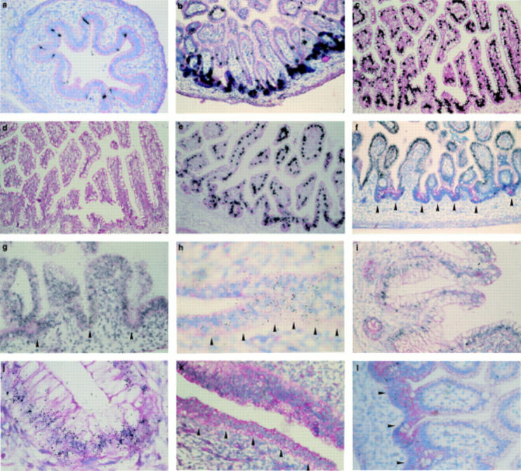
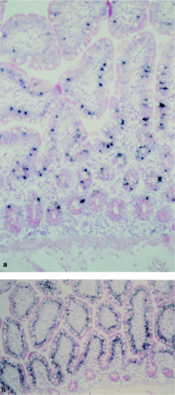
Aims: To assess whether the expression of mucin genes in the intestinal tract is linked to the stage of cellular differentiation and tissue development, by studying the expression of six mucin genes in human fetal small intestine and colon, and also adult tissues.

Methods: In situ hybridisation was used to study mRNA expression of MUC2, MUC3, MUC4, MUC5B, MUC5AC, and MUC6 in 32 human embryos and fetuses (6.5-27 weeks gestation). Normal adult mucosae were used as controls.

Results: Three mucin genes, MUC2, MUC4, and MUC5AC, were differently expressed in fetal intestine compared with expression in normal adults.

Conclusion: These differences in mucin gene expression suggest a possible regulatory role for these products in intestinal epithelial cell differentiation.


Figures

Figure 1
Figure 1
In situ hybridisation for mucin gene mRNAs in embryonic and fetal intestine. (a) Small intestine at 10 weeks gestation with MUC2 probe: the signal is located predominantly between the primordial villi (methyl green pyronin counterstain; original magnification ×200). (b) Small intestine at 12 weeks' gestation with MUC2 probe: the signal is located predominantly within immature crypts of Lieberkühn (methyl green pyronin counterstain; original magnification ×200). (c, d) Ileum at 23 weeks' gestation: (c) with MUC2 probe: signal is stronger in the crypts, but the majority of villous goblet cells are labelled; (d) with MUC2 probe and an excess of cold unlabelled MUC2 oligonucleotide (negative control): hybridisation is negative (methyl green pyronin counterstain; original magnification ×200). (e) Ileum at 26 weeks' gestation with MUC2 probe: signal is distributed in goblet cells both on villi and in crypts (methyl green pyronin counterstain; original magnification ×200). (f) Small intestine at 12 weeks' gestation with MUC3 probe: continuous and homogeneous labelling along the villous epithelium, both in goblet and absorptive cells; no labelling present in crypts (arrows) (methyl green pyronin counterstain; original magnification ×400). (g) Small intestine at 14.1 weeks' gestation with MUC3 probe: continuous and homogeneous labelling along the villous epithelium, both in goblet and absorptive cells; weak labelling is also present in crypts (arrows) (methyl green pyronin counterstain; original magnification ×500). (h) Anterior portion of primitive gut at 6.5 weeks' gestation with MUC4 probe: continuous labelling along epithelium (methyl green pyronin counterstain; original magnification ×1000). (i, j) Colon at 23 weeks' gestation with MUC4 probe: signal is located in the perinuclear region of goblet cells (methyl green pyronin counterstain; original magnification: (i) ×400; (j) ×1000). (k) Middle portion of primitive gut at 8 weeks' gestation with MUC5AC probe: continuous labelling along the undifferentiated epithelium (methyl green pyronin counterstain; original magnification ×500). (l) Ileum at 12 weeks' gestation with MUC5AC probe: labelling is limited to clusters of epithelial cells both on villi and in crypts (methyl green pyronin counterstain; original magnification ×500).
Figure 2
Figure 2
In situ hybridisation for mucin gene mRNAs in adult intestine. (a) Jejunum with MUC2 probe: signal is located in the perinuclear region of goblet cells both on villi and in crypts (methyl green pyronin counterstain; original magnification ×200). (b) Jejunum with MUC3 probe: continuous and homogeneous labelling along the villous epithelium, both in goblet and absorptive cells (methyl green pyronin counterstain; original magnification ×200).

References

    1. Am J Respir Cell Mol Biol. 1991 Aug;5(2):178-85 - PubMed
    1. Proc Natl Acad Sci U S A. 1988 Apr;85(7):2320-3 - PubMed
    1. Semin Cancer Biol. 1991 Dec;2(6):389-400 - PubMed
    1. Cancer Res. 1993 Feb 1;53(3):641-51 - PubMed
    1. J Biol Chem. 1993 Mar 15;268(8):5879-85 - PubMed

Publication types

MeSH terms