

Case Reports
doi: 10.1136/gut.43.4.575.
Abscopal regression of hepatocellular carcinoma after radiotherapy for bone metastasis
Affiliations
- PMID: 9824589
- PMCID: PMC1727260
- DOI: 10.1136/gut.43.4.575
Item in Clipboard
Case Reports
Abscopal regression of hepatocellular carcinoma after radiotherapy for bone metastasis
Gut.
1998 Oct.
Abstract
Spontaneous regression of hepatocellular carcinoma is a rare phenomenon. Abscopal regression of tumours resulting from the effect of irradiation of a tissue on a remote non-irradiated tissue is also rare. The case of a 76 year old Japanese man with hepatocellular carcinoma that regressed after radiotherapy for thoracic vertebral bone metastasis is described. Serum levels of tumour necrosis factor-alpha increased after radiotherapy. The findings suggests that such abscopal related regression may be associated with host immune response, involving cytokines such as tumour necrosis factor-alpha.
Figures
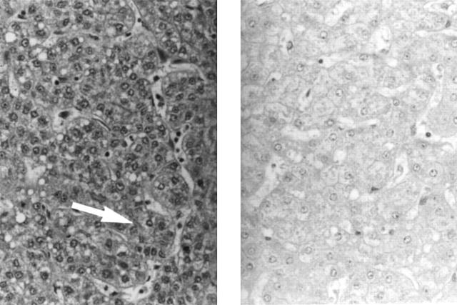
Histological examination of a section of the tumour of
the liver showing a well differentiated hepatocellular carcinoma (left
panel). Note the rather slender cell cord composed of small tumour
cells. Microacinar formation (arrow) is also seen. Nuclear crowding is
evident compared with that in control tissue obtained from the same
liver (right panel). Haematoxylin and eosin; original magnification
×80.

Hepatic angiogram obtained in November 1992 showing
multiple hypervascular stains in the right lobe (arrows).
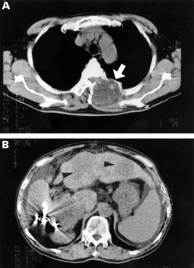
Thoracic and abdominal computed tomography scans
obtained in June 1995 showing a mass in the second thoracic vertebra
(arrow) (A) and multiple low densities in the liver (arrowheads) (B).
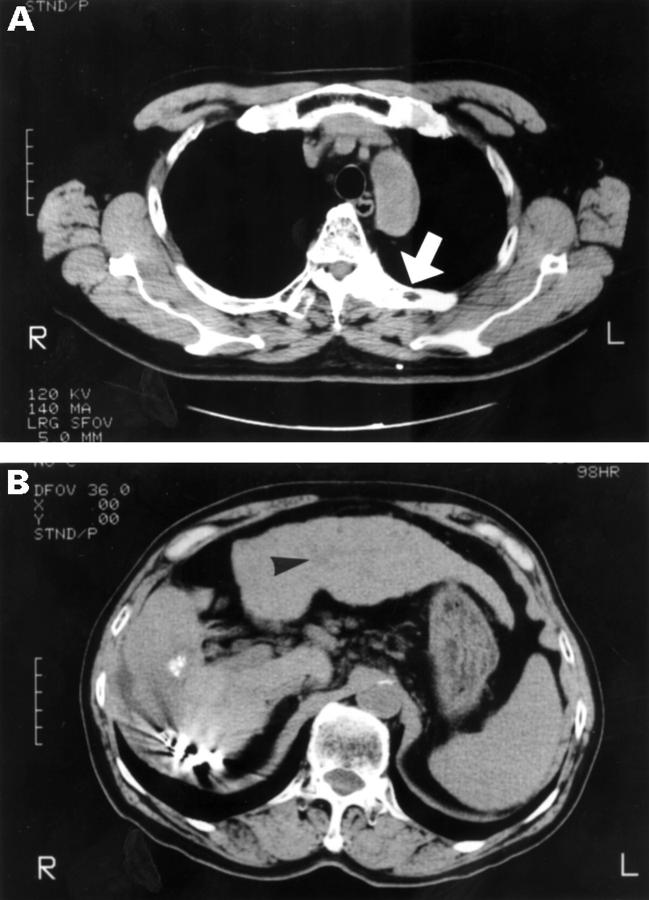
Thoracic and abdominal computed tomography scans
showing disappearance of the mass from the second thoracic vertebra
(arrow) (A) and a remarkable regression of the hepatic lesions
(arrowhead) (B) one month and 10 months after radiotherapy
respectively.
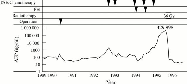
Clinical course. Note the immediate fall in serum α fetoprotein (AFP) and increase in tumour necrosis factor (TNF)-α
after radiotherapy for bone metastasis. TAE, transcatheter arterial
embolisation; PEI, percutaneous ethanol injection; HGF, hepatocyte
growth factor.
Publication types
MeSH terms
LinkOut - more resources
Full Text Sources
Other Literature Sources
Medical