. 1998 Dec 15;513 ( Pt 3)(Pt 3):819-33.
doi: 10.1111/j.1469-7793.1998.819ba.x.

Effects of mitochondrial uncouplers on intracellular calcium, pH and membrane potential in rat carotid body type I cells

Affiliations

Effects of mitochondrial uncouplers on intracellular calcium, pH and membrane potential in rat carotid body type I cells

K J Buckler et al. J Physiol. .

Abstract

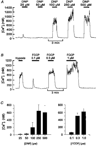
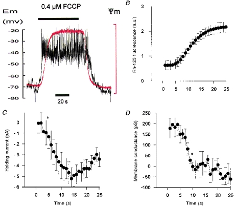
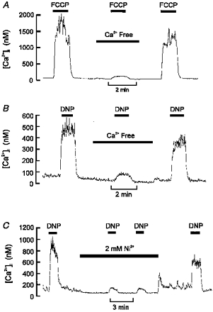
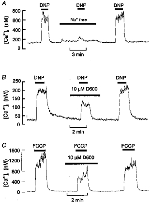
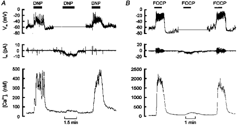
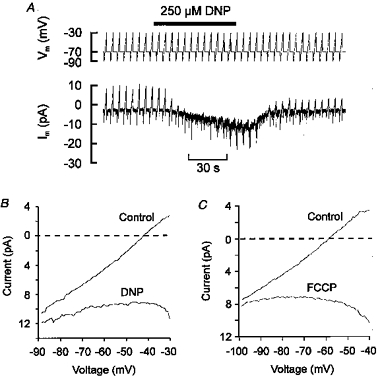
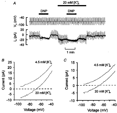
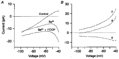
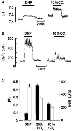
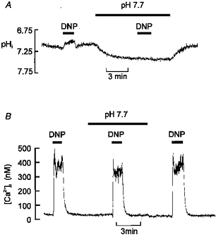
1. Mitochondrial uncouplers are potent stimulants of the carotid body. We have therefore investigated their effects upon isolated type I cells. Both 2,4-dinitrophenol (DNP) and carbonyl cyanide p-trifluoromethoxyphenyl hydrazone (FCCP) caused an increase in [Ca2+]i which was largely inhibited by removal of extracellular Ca2+ or Na+, or by the addition of 2 mM Ni2+. Methoxyverapamil (D600) also partially inhibited the [Ca2+]i response. 2. In perforated-patch recordings, the rise in [Ca2+]i coincided with membrane depolarization and was greatly reduced by voltage clamping the cell to -70 mV. Uncouplers also inhibited a background K+ current and induced a small inward current. 3. Uncouplers reduced pHi by 0.1 unit. Alkaline media diminished this acidification but had no effect on the [Ca2+]i response. 4. FCCP and DNP also depolarized type I cell mitochondria. The onset of mitochondrial depolarization preceded changes in cell membrane conductance by 3-4 s. 5. We conclude that uncouplers excite the carotid body by inhibiting a background K+ conductance and inducing a small inward current, both of which lead to membrane depolarization and voltage-gated Ca2+ entry. These effects are unlikely to be caused by cell acidification. The inhibition of background K+ current may be related to the uncoupling of oxidative phosphorylation.


Figures

Figure 1
Figure 1. Effects of uncouplers on [Ca2+]i in type I cells
Recordings of [Ca2+]i in individual type I cells and their responses to graded levels of DNP (A) or FCCP (B). Data in B also include a control response to a hypoxic (PO2, ∼5 Torr) stimulus for comparison. C, mean [Ca2+]i responses to graded levels of DNP (n = 3 at 25 μm, n = 5 all other DNP data) and FCCP (n = 6). Error bars indicate s.e.m.
Figure 10
Figure 10. Effects of FCCP on membrane properties and mitochondrial potential
A, simultaneous recording of membrane potential (Vm, black trace) and mitochondrial membrane potential (Ψm, red trace, note that upward deflection equals depolarization) during application of 0.4 μm FCCP. B, C and D, time course of change in mitochondrial potential (Rh-123 fluorescence in arbitrary units, a.u.), holding current and membrane conductance, respectively, following application of 0.4 μm FCCP. Mean ± s.e.m. values from five cells. Asterisks indicate the first data point that is significantly different (P < 0.05) from the control level (control equals data obtained in the first second, i.e. just before FCCP actually reaches the recording chamber). All data were recorded simultaneously in cells voltage clamped at −70 mV and subjected to 500 ms voltage ramps from −100 to −50 mV at 1 Hz. Membrane conductance was calculated by fitting a linear regression over the range −60 to −50 mV). Electrophysiological recordings were performed using the perforated-patch technique with pipette filling solution C.
Figure 2
Figure 2. Uncoupler-evoked rise in [Ca2+]i is dependent upon Ca2+ influx
A, effect of removal of extracellular Ca2+ (+ 1 mm EGTA) on [Ca2+]i response to 0.3 μm FCCP. B, effect of removal of extracellular Ca2+ on [Ca2+]i response to 250 μm DNP. C, effect of 2 mm Ni2+ on [Ca2+]i response to 250 μm DNP. Note that both Ca2+ removal and 2 mm Ni2+ substantially inhibited the [Ca2+]i response to uncouplers. A small residual rise in [Ca2+]i remained in most cells.
Figure 3
Figure 3. Effects of Na+ removal and Ca2+ channel antagonists on [Ca2+]i response to uncouplers
A, effects of removal of extracellular Na+ (replaced with NMDG) on the [Ca2+]i response to 250 μm DNP. B and C, effects of 10 μm D600 on the [Ca2+]i response to 250 μm DNP (B) or 0.3 μm FCCP (C). Note that both removal of extracellular Na+ and addition of D600 inhibited the response to uncoupler (see text for statistics).
Figure 4
Figure 4. Effects of uncouplers on membrane potential and [Ca2+]i
A and B, upper panels show recordings of membrane potential (perforated-patch technique), middle panels membrane current and lower panels [Ca2+]i recorded simultaneously in single type I cells. Each experiment started and finished in current-clamp mode (Im, 0) and shows both a membrane depolarization and a rise in [Ca2+]i in response to uncoupler (250 μm DNP in A, 0.3 μm FCCP in B). In the middle section of each experiment (corresponding to the flat portion of the membrane potential trace) the cells were voltage clamped to −60 (A) or −70 mV (B). Under voltage-clamp conditions DNP and FCCP evoked an inward shift in holding current, consistent with the depolarization seen under current-clamp conditions, and a greatly attenuated rise in [Ca2+]i. Pipette filling solutions A (A) and C (B).
Figure 5
Figure 5. Uncouplers reduce membrane conductance
A, a single type I cell voltage clamped at −70 mV and subjected to repetitive voltage ramps (2 s duration) from −90 to −30 mV every 5 s. Pipette filling solution A. Note that DNP caused both an inward shift in holding current and a decline in the amplitude of the ramp current. B, mean current-voltage relationship (n = 4) obtained using the above protocol both under control conditions and in the presence of DNP. C, mean current-voltage relationship (n = 8) obtained using 500 ms voltage ramps from −100 to −40 mV applied every 5 s under control conditions and in the presence of 0.3 μm FCCP. Pipette filling solution C. Note: (a) that membrane conductance is greatly reduced in the presence of either DNP or FCCP; (b) that a zero current (resting) potential does not exist in this voltage range in the presence of DNP or FCCP; and (c) an inwardly rectifying current in the presence of DNP at potentials below −40 mV.
Figure 6
Figure 6. Current-voltage relationship of uncoupler-sensitive current
A, type I cell voltage clamped at −70 mV and subjected to 500 ms voltage ramps from −100 to −40 mV at two levels of extracellular K+ in the presence and absence of 250 μm DNP. Pipette filling solution B. B and C, mean current-voltage relationships of DNP (B)- and FCCP (C)-sensitive current obtained by subtracting the I-V relationship obtained in the presence of uncoupler from the control I-V relationship. Note the marked rightward and downward (depolarizing) shift in the uncoupler-sensitive current in high [K+]o. Note also the clear reversal potentials obtained in 20 mm[K+]o with both uncouplers. DNP, n = 8. FCCP, n = 8, pipette filling solution C.
Figure 7
Figure 7. FCCP induces an inward current
A, mean current-voltage relationship (n = 5) constructed from voltage ramps (−100 to −40 mV) applied every 5 s from a holding potential of −70 mV. Pipette filling solution C. I-V curves for control conditions and those obtained in the presence of 5 mm Ba2+ (no Ca2+) and 5 mm Ba2++ 0.3 μm FCCP are shown. B, difference currents from data in A. Ba2+-sensitive background K+ current (control minus Ba2+) (a). FCCP-induced current (Ba2++ FCCP minus Ba2+) (b). Combined FCCP-induced inward current and Ba2+-sensitive K+ current (control minus Ba2++ FCCP) (c). Note that the FCCP-induced current changed little over the voltage range shown.
Figure 8
Figure 8. Effects of DNP on pHi in comparison with other acidic stimuli
A, effects of 250 μm DNP and hypercapnic acidosis (10 % CO2, pHo 7.15) on pHi. Note that although DNP evokes a rapid fall in pHi the decrease is small in comparison to the effects of hypercapnic acidosis. B, effects of DNP and 10 % CO2 on [Ca2+]i in another cell. C, comparison of the effect of DNP upon both pHi and [Ca2+]i with those of 10 and 20 % CO2. Filled bars represent fall in pHi, open bars rise in [Ca2+]i (CO2 data from Buckler et al. 1991a; Buckler & Vaughan-Jones, 1993a).
Figure 9
Figure 9. Effects of extracellular alkalosis on pHi and [Ca2+]i responses to DNP
A, effects of increasing extracellular pH upon resting pHi and the response to DNP. pHo was elevated to 7.7 by doubling HCO3 (to 46 mm) with an equimolar reduction in NaCl. Note that the alkaline bathing medium increases pHi and reduces the acidification caused by DNP so that pHi remains alkaline compared with the control level. B, effects of the above alkalinizing protocol on the [Ca2+]i response to DNP. Note that pre-alkalinization does not prevent the [Ca2+]i response to DNP.

References

    1. Alekseev AE, Gomez LA, Aleksandrova LA, Brady PA, Terzic A. Opening of cardiac sarcolemmal KATP channels by dinitrophenol separate from metabolic inhibition. Journal of Membrane Biology. 1997;157:203–214. . - DOI - PubMed
    1. Anichcov SV, Belen'kii ML. Pharmacology of the Carotid Body Chemoreceptors. New York: Macmillan Publishing; 1963.
    1. Biscoe TJ, Duchen MR. Responses of type I cells dissociated from the rabbit carotid body to hypoxia. The Journal of Physiology. 1990a;428:39–59. - PMC - PubMed
    1. Biscoe TJ, Duchen MR. The cellular basis of transduction in carotid chemoreceptors. American Journal of Physiology. 1990b;258:L271–278. - PubMed
    1. Boron WF, De Weer P. Intracellular pH transients in squid giant axons caused by CO2, NH3, and metabolic inhibitors. Journal of General Physiology. 1976;67:91–112. - PMC - PubMed

Publication types

MeSH terms

LinkOut - more resources