. 1998 Dec 15;513 ( Pt 3)(Pt 3):835-44.
doi: 10.1111/j.1469-7793.1998.835ba.x.

Apamin- and nitric oxide-sensitive biphasic non-adrenergic non-cholinergic inhibitory junction potentials in the rat anococcygeus muscle

Affiliations

Apamin- and nitric oxide-sensitive biphasic non-adrenergic non-cholinergic inhibitory junction potentials in the rat anococcygeus muscle

S Selemidis et al. J Physiol. .

Abstract

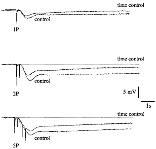
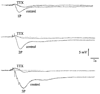
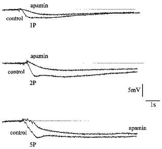
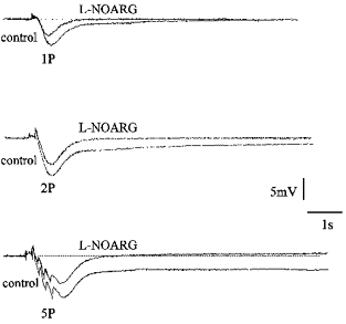
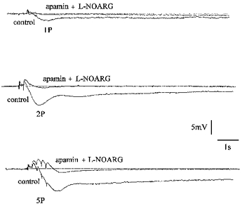
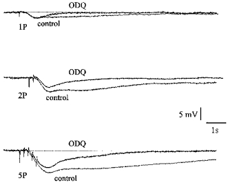
1. Changes in membrane potential following electrical field stimulation (EFS; 1, 2 and 5 pulses at 5 Hz, 0.5 ms duration, 60-80 V) of non-adrenergic non-cholinergic (NANC) inhibitory nerves in the rat isolated anococcygeus muscle were measured using standard intracellular recording techniques. Resting membrane potential ranged between -60 and -70 mV. 2. In the presence of guanethidine (30 microM), atropine (1 microM), propranolol (1 microM) and phentolamine (0.05 microM) to establish NANC conditions, the membrane potential depolarized to between -40 and -50 mV. Under these conditions, EFS caused pulse-dependent, tetrodotoxin (1 microM)-sensitive biphasic inhibitory junction potentials (IJPs) comprising a fast onset and time-to-peak phase followed by a second, slower phase that delayed repolarization. The duration of NANC IJPs ranged between 10 and 20 s. 3. Inhibition of small-conductance Ca2+-activated K+ channels with apamin (0.1 microM) selectively blocked the first fast phase of the NANC IJP, whereas inhibitors of large-conductance Ca2+-activated K+ channels (charybdotoxin and iberiotoxin) and ATP-sensitive K+ channels (glibenclamide) all had no effect on NANC IJPs. 4. Both the nitric oxide synthase inhibitor N G-nitro-L-arginine (L-NOARG; 100 microM) and the inhibitor of soluble guanylate cyclase 1-H-oxodiazol-[1,2,4]-[4,3-a] quinoxaline-1-one (ODQ; 10 microM) had no effect on the first fast phase of the NANC IJP. Each treatment, however, markedly inhibited the slow phase with the duration of the IJP reduced to between 1 and 3 s. The L-NOARG-resistant fast phase of the NANC IJP was almost abolished by the subsequent addition of apamin (0.1 microM). 5. In conclusion, the present study demonstrates unequivocal NANC nerve-mediated biphasic IJPs in the rat isolated anococcygeus. We propose that nitric oxide (NO), via activation of cGMP-dependent K+ channels, and a non-NO inhibitory factor which activates apamin-sensitive K+ channels contribute to NANC nerve-evoked IJPs in the rat anococcygeus.


Figures

Figure 1
Figure 1. Effect of time on NANC IJPs
Superimposed digitized recordings of membrane potential showing the 1st control (control) and sequential, time control (30 min later) inhibitory junction potentials (IJPs) following electrical field stimulation (1, 2 and 5 pulses (P) at 5 Hz every 45–60 s, 0.5 ms duration, 60–80 V) of NANC inhibitory nerves in rat isolated anococcygeus muscle. A downward deflection signifies hyperpolarization whereas transient spikes indicate stimulus artefacts for each pulse. The amplitude of the IJPs in time controls was slightly reduced compared with the 1st control IJPs; however, the duration and time to peak remained unaffected. Horizontal dotted line indicates the pre-stimulus resting membrane potential (−40 to −50 mV). Vertical (mV) and horizontal (time scale; s) bars apply to all traces. All recordings shown in this panel are from a single cell and are representative of 6 cells from 6 separate animals.
Figure 2
Figure 2. Effect of TTX on NANC IJPs
Superimposed digitized recordings of membrane potential showing the 1st control (control) inhibitory junction potentials (IJPs) and sequential IJPs in the presence of tetrodotoxin (TTX; 1 μm) following electrical field stimulation (1, 2 and 5 pulses (P) at 5 Hz every 45–60 s, 0.5 ms duration, 60–80 V) of NANC inhibitory nerves in rat isolated anococcygeus muscle. A downward deflection signifies hyperpolarization whereas transient spikes indicate stimulus artefacts for each pulse. TTX almost abolished the IJPs but in some cases (as shown here) it revealed a small depolarization followed by a small hyperpolarization. Horizontal dotted line indicates the pre-stimulus resting membrane potential (−40 mV). Vertical (mV) and horizontal (time scale; s) bars apply to all traces. All recordings shown in this panel are from a single cell and are representative of 6 cells from 6 separate animals.
Figure 3
Figure 3. Effect of apamin on NANC IJPs
Superimposed digitized recordings of membrane potential showing the 1st control (control) inhibitory junction potentials (IJPs) and sequential IJPs in the presence of apamin (0.1 μm) following electrical field stimulation (1, 2 and 5 pulses (P) at 5 Hz every 45–60 s, 0.5 ms duration, 60–80 V) of NANC inhibitory nerves in rat isolated anococcygeus muscle. A downward deflection signifies hyperpolarization. Apamin inhibited the fast phase of the IJP by reducing the peak amplitude and by increasing the time to peak. Horizontal dotted line indicates the pre-stimulus resting membrane potential (−48 to −52 mV). Vertical (mV) and horizontal (time scale; s) bars apply to all traces. All recordings shown in this panel are from a single cell and are representative of 6 cells from 6 separate animals.
Figure 4
Figure 4. Effect of NG-nitro-L-arginine (l-NOARG) on NANC IJPs
Superimposed digitized recordings of membrane potential showing 1st control (control) inhibitory junction potentials (IJPs) and sequential IJPs in the presence of l-NOARG (100 μm) following electrical field stimulation (1, 2 and 5 pulses (P) at 5 Hz every 45–60 s, 0.5 ms duration, 60–80 V) of NANC inhibitory nerves in rat isolated anococcygeus muscle. Downward deflection signifies hyperpolarization whereas transient spikes indicate stimulus artefacts for each pulse. Note the time to peak of the IJP was unaffected by l-NOARG treatment but the duration reduced to between 1 and 3 s, therefore, l-NOARG had no effect on the fast phase (compared with time controls in Fig. 1) but abolished the second phase. Horizontal dotted line indicates the pre-stimulus resting membrane potential (−40 to −50 mV). Vertical (mV) and horizontal (time scale; s) bars apply to all traces. All recordings shown in this panel are from a single cell and are representative of 6 cells from 6 separate animals.
Figure 5
Figure 5. Effect of apamin and l-NOARG in combination on NANC IJPs
Superimposed digitized recordings of membrane potential showing 1st control (control) inhibitory junction potentials (IJPs) and sequential IJPs in the presence of a combination of apamin (0.1 μm) and l-NOARG (100 μm) following electrical field stimulation (1, 2 and 5 pulses (P) at 5 Hz every 45–60 s, 0.5 ms duration, 60–80 V) of NANC inhibitory nerves in rat isolated anococcygeus muscle. A downward deflection signifies hyperpolarization whereas transient spikes indicate stimulus artefacts for each pulse. The combined treatment of apamin and l-NOARG nearly abolished the IJP and in some cases revealed a small depolarization which is most likely to be a stimulus artefact. Horizontal dotted line indicates the pre-stimulus resting membrane potential (−40 to −45 mV). Vertical (mV) and horizontal (time scale; s) bars apply to all traces. All recordings shown in this panel are from a single cell and are representative of 6 cells from 6 separate animals.
Figure 6
Figure 6. Effect of ODQ on NANC IJPs
Superimposed digitized recordings of membrane potential showing the 1st control (control) inhibitory junction potentials (IJPs) and sequential IJPs in the presence of ODQ (10 μm) following electrical field stimulation (1, 2 and 5 pulses (P) at 5 Hz every 45–60 s, 0.5 ms duration, 60–80 V) of NANC inhibitory nerves in rat isolated anococcygeus muscle. A downward deflection signifies hyperpolarization whereas transient spikes indicate stimulus artefacts for each pulse. Like l-NOARG (Fig. 4), ODQ inhibited the second, slow phase of the IJP because the time to peak was unaffected and the duration reduced to between 1 and 3 s. Horizontal dotted line indicates the pre-stimulus resting membrane potential (−45 mV). Vertical (mV) and horizontal (time scale; s) bars apply to all traces. All recordings shown in this panel are from a single cell and are representative of 6 cells from 6 separate animals.

Similar articles

References

    1. Bennett MA. Non-adrenergic non-cholinergic (NANC) transmission to smooth muscle: 35 years on. Progress in Neurobiology. 1997;52:159–195. - PubMed
    1. Bolotina VM, Najibi S, Palacino JJ, Pagano PJ, Cohen RA. Nitric oxide directly activates calcium-dependent potassium channels in vascular smooth muscle. Nature. 1994;368:850–853. - PubMed
    1. Bridgewater M, Cunnane TC, Brading AF. Characteristic features of inhibitory junction potentials evoked by single stimuli in the guinea-pig isolated taenia caeci. The Journal of Physiology. 1995;485:145–155. - PMC - PubMed
    1. Burnstock G. Purinergic nerves. Pharmacological Reviews. 1972;24:509–581. - PubMed
    1. Burnstock G. Overview: purinergic mechanisms. Annals of the New York Academy of Sciences. 1990;603:1–18. - PubMed

Publication types

MeSH terms

LinkOut - more resources