Comparative Study
. 1998 Dec;66(12):5643-9.
doi: 10.1128/IAI.66.12.5643-5649.1998.

Identification of immunodominant regions within the C-terminal cell binding domain of intimin alpha and intimin beta from enteropathogenic Escherichia coli

Affiliations
Comparative Study

Identification of immunodominant regions within the C-terminal cell binding domain of intimin alpha and intimin beta from enteropathogenic Escherichia coli

J Adu-Bobie et al. Infect Immun. 1998 Dec.

Abstract

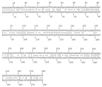
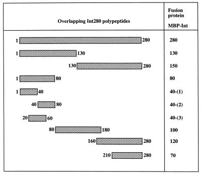
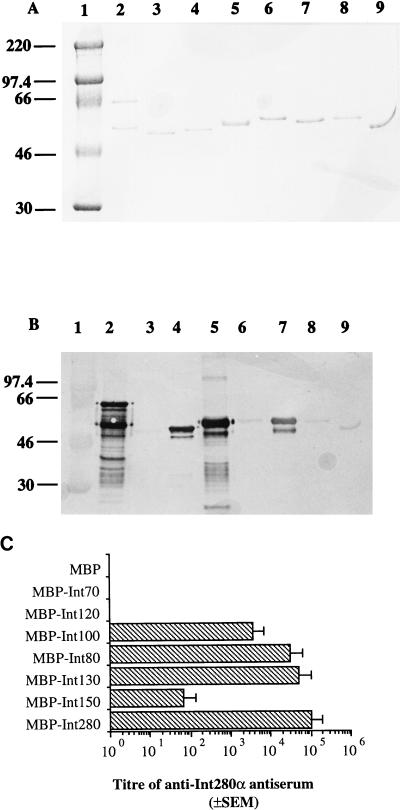
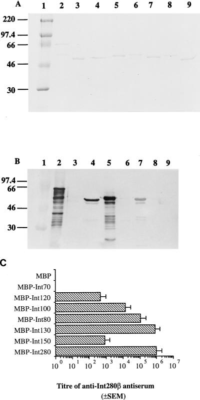
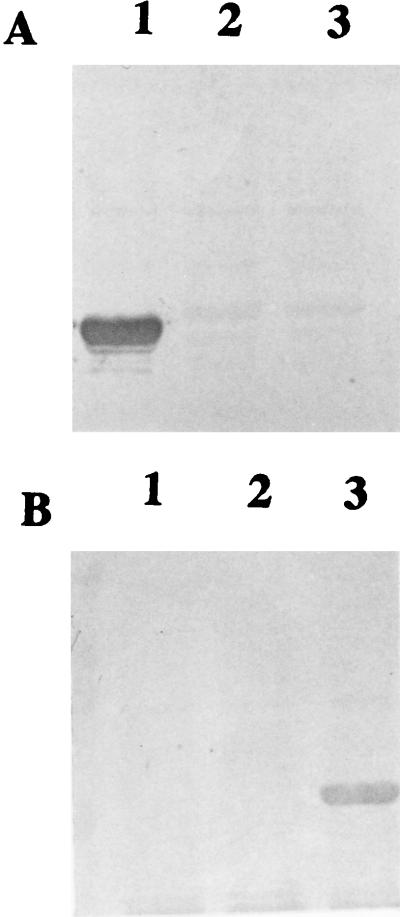
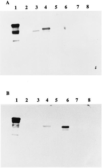
Enteropathogenic Escherichia coli (EPEC) strains are a common cause of infantile diarrhea in developing countries. EPEC strains induce a characteristic attaching and effacing (A/E) lesion on epithelial cells. A/E lesion formation requires intimin, an outer membrane adhesin protein. The cell-binding activity of intimin is localized at the C-terminal 280 amino acids of the polypeptide (Int280). So far, four distinct Int280 types (alpha, beta, gamma, and delta) have been identified. The aim of this study was to identify immunodominant regions within the Int280alpha and Int280beta domains. Recombinant DNA was used to construct and express overlapping polypeptides spanning these domains. Rabbit anti-Int280 antisera and human colostral immunoglobulin A were reacted with these polypeptides in Western blots and enzyme-linked immunosorbent assays. The results obtained with the rabbit antisera showed the presence of two separate immunodominant regions which are common to both Int280alpha and Int280beta. The first localized within the N-terminal region of Int280, and the second localized between amino acids 80 and 130. The results with the human colostra revealed one reactivity pattern against the Int280alpha fragments but two different reactivity patterns against the Int280beta domain.


Figures

FIG. 1
FIG. 1
Alignment of the amino acid sequence of Int280α from the EPEC E2348/69 domain (upper row) and Int280β from the EPEC O114:H2 domain (lower row). Dots represent identical residues. Lines represent conserved substitutions.
FIG. 2
FIG. 2
Schematic representation of the overlapping MBP-Int280-derived polypeptides. The numbers on both sides of the fragments mark the first and last amino acids of each fragment within the Int280 domain.
FIG. 3
FIG. 3
Coomassie blue staining of SDS-PAGE gel (A) and immunoblotting (B) and ELISA (C) of anti-Int280α antiserum with Int280α-derived fusion proteins. Strong reactivity on the Western blot is observed with Int280 (B, lane 2), Int80 (B, lane 4), Int130 (B, lane 5), and Int100 (B, lane 7). Low reactivity is observed with Int150 (B, lane 6), Int120 (B, lane 8), and Int70 (B, lane 9). No reactivity is seen with MBP (lane 3). The electrophoretic migration of molecular mass markers (in kilodaltons) is shown in lane 1 (A and B). The reactivity of Int280α antiserum with the different overlapping Int280α-derived fusion proteins in the ELISA produced results in agreement with those of the Western blot (C). High antibody titers were observed for Int280, -130, -80, and -100, and a low titer was observed for Int150. No reactivity was seen for Int120, Int70, or MBP.
FIG. 4
FIG. 4
Coomassie blue staining of SDS-PAGE gel (A) and immunoblotting (B) and ELISA (C) of the reactivity of anti-Int280β antiserum (1:10,000 dilution) with the MBP-Int280β-derived fusion proteins. Strong reactivity on the Western blot is observed with Int280 (B, lane 2), Int80 (B, lane 4), Int130 (B, lane 5), and Int100 (B, lane 7). Low reactivity is seen with Int150 (B, lane 6). No reactivity is seen with Int120 (B, lane 8), Int70 (B, lane 9), or MBP (B, lane 3). MBP-Int280 preparations consistently resulted in doublet bands; this appears to represent proteolytic sensitivity introduced into the MBP part of the fusion protein (15). Molecular mass markers (A and B, lane 1) are given in kilodaltons. Analysis of the Int280β antiserum with the different overlapping Int280b-derived fusion protein ELISA (C) revealed that high antibody titers were observed for Int280, -130, -80, and -100, while lower titers were observed against Int150 and Int120. No reactivity was seen for Int70 or MBP.
FIG. 5
FIG. 5
Reactivity of Int280α and Int280β antisera with Int80-derived overlapping polypeptides. Anti-Int280α antiserum reacted with Int40-(1)α (A, lane 1) but showed no reactivity against Int40-(2)α or Int40-(3)α (A, lanes 2 and 3, respectively). In contrast anti-Int280β antiserum reacted with Int40-(3)β (B, lane 3) and showed no reactivity with Int40-(1)β or Int40-(2)β (B, lanes 1 and 2, respectively).
FIG. 6
FIG. 6
Reactivity of colostrum samples with MBP-Int fusion proteins. Reaction of the four individual colostra with Int280α-derived MBP fusion protein revealed a single reactivity pattern (A) in which the IgA antibodies reacted strongly with Int280α (lane 1), moderately with Int130α (lane 4), and weakly with Int80α (lane 3). No reactivity was seen with Int150α, Int120α, Int70α, or MBP (A, lanes 5, 7, 8, and 2, respectively). Reaction of the same four colostrum samples with Int280β-derived fusion proteins revealed, in two samples, the same reactivity pattern as that seen with reaction against the Int280α-derived polypeptides (data not shown), while the other two samples reacted strongly with Int280β (B, lane 1), moderately with Int100β (B, lane 6), and weakly with Int130β (B, lane 4). No reactivity was seen with Int80, Int150, Int120, Int70, or MBP (B, lanes 3, 5, 7, 8, and 2, respectively).

References

    1. Adu-Bobie J, Frankel G, Bain C, Goncalves A Z, Trabulsi L R, Douce G, Knutton S, Dougan G. Detection of intimins α, β, γ, and δ, four intimin derivatives expressed by attaching and effacing microbial pathogens. J Clin Microbiol. 1998;36:662–668. - PMC - PubMed
    1. Agin T S, Wolf M K. Identification of a family of intimins common to Escherichia coli causing attaching-effacing lesions in rabbits, humans, and swine. Infect Immun. 1997;65:320–326. - PMC - PubMed
    1. Burnette W N. “Western blotting”: electrophoretic transfer of proteins from sodium dodecyl sulphate-polyacrylamide gels to unmodified nitrocellulose and radiographic detection with antibody and radioiodinated protein A. Anal Biochem. 1981;112:195–203. - PubMed
    1. Camara L M, Carbonare S B, Silva M L, Carneiro Sampaio M M. Inhibition of enteropathogenic Escherichia coli (EPEC) adhesion to HeLa cells by human colostrum: detection of specific IgA related to EPEC outer-membrane proteins. Int Arch Allergy Immunol. 1994;103:307–310. - PubMed
    1. Cantey J R, Blake R K. Diarrhea due to Escherichia coli in rabbit: a novel mechanism. J Infect Dis. 1977;135:454–462. - PubMed

Publication types

MeSH terms

LinkOut - more resources