Cloning, expression, and sequencing of a cell surface antigen containing a leucine-rich repeat motif from Bacteroides forsythus ATCC 43037
- PMID: 9826345
- PMCID: PMC108721
- DOI: 10.1128/IAI.66.12.5703-5710.1998
Cloning, expression, and sequencing of a cell surface antigen containing a leucine-rich repeat motif from Bacteroides forsythus ATCC 43037
Abstract
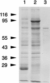
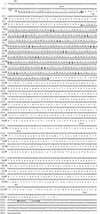
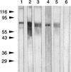
Bacteroides forsythus is a recently recognized human periodontopathogen associated with advanced, as well as recurrent, periodontitis. However, very little is known about the mechanism of pathogenesis of this organism. The present study was undertaken to identify the surface molecules of this bacterium that may play roles in its adherence to oral tissues or triggering of a host immune response(s). The gene (bspA) encoding a cell surface-associated protein of B. forsythus with an apparent molecular mass of 98 kDa was isolated by immunoscreening of a B. forsythus gene library constructed in a lambda ZAP II vector. The encoded 98-kDa protein (BspA) contains 14 complete repeats of 23 amino acid residues that show partial homology to leucine-rich repeat motifs. A recombinant protein containing the repeat region was expressed in Escherichia coli, purified, and utilized for antibody production, as well as in vitro binding studies. The purified recombinant protein bound strongly to fibronectin and fibrinogen in a dose-dependent manner and further inhibited the binding of B. forsythus cells to these extracellular matrix (ECM) components. In addition, adult patients with B. forsythus-associated periodontitis expressed specific antibodies against the BspA protein. We report here the cloning and expression of an immunogenic cell surface-associated protein (BspA) of B. forsythus and speculate that it mediates the binding of bacteria to ECM components and clotting factors (fibronectin and fibrinogen, respectively), which may be important in the colonization of the oral cavity by this bacterium and is also a target for the host immune response.
Figures


Similar articles
-
Development of a gene inactivation system for Bacteroides forsythus: construction and characterization of a BspA mutant.Infect Immun. 2001 Jul;69(7):4686-90. doi: 10.1128/IAI.69.7.4686-4690.2001. Infect Immun. 2001. PMID: 11402017 Free PMC article.
-
[Cloning and sequence analysis of a sialidase gene (siaHI) from Bacteroides forsythus ATCC 43037].Kokubyo Gakkai Zasshi. 2002 Jun;69(2):119-27. doi: 10.5357/koubyou.69.119. Kokubyo Gakkai Zasshi. 2002. PMID: 12136659 Japanese.
-
Tannerella forsythia-induced alveolar bone loss in mice involves leucine-rich-repeat BspA protein.J Dent Res. 2005 May;84(5):462-7. doi: 10.1177/154405910508400512. J Dent Res. 2005. PMID: 15840784
-
Multiple functions of the leucine-rich repeat protein LrrA of Treponema denticola.Infect Immun. 2004 Aug;72(8):4619-27. doi: 10.1128/IAI.72.8.4619-4627.2004. Infect Immun. 2004. PMID: 15271922 Free PMC article.
-
Applications of blood group antigen expression systems for antibody detection and identification.Transfusion. 2007 Jul;47(1 Suppl):85S-8S. doi: 10.1111/j.1537-2995.2007.01317.x. Transfusion. 2007. PMID: 17593293 Review. No abstract available.
Cited by
-
Taxon-Specific Proteins of the Pathogenic Entamoeba Species E. histolytica and E. nuttalli.Front Cell Infect Microbiol. 2021 Mar 19;11:641472. doi: 10.3389/fcimb.2021.641472. eCollection 2021. Front Cell Infect Microbiol. 2021. PMID: 33816346 Free PMC article. Review.
-
Single cell genomics of uncultured, health-associated Tannerella BU063 (Oral Taxon 286) and comparison to the closely related pathogen Tannerella forsythia.PLoS One. 2014 Feb 14;9(2):e89398. doi: 10.1371/journal.pone.0089398. eCollection 2014. PLoS One. 2014. PMID: 24551246 Free PMC article.
-
Population-Genomic Insights into Variation in Prevotella intermedia and Prevotella nigrescens Isolates and Its Association with Periodontal Disease.Front Cell Infect Microbiol. 2017 Sep 21;7:409. doi: 10.3389/fcimb.2017.00409. eCollection 2017. Front Cell Infect Microbiol. 2017. PMID: 28983469 Free PMC article.
-
Characterization and scope of S-layer protein O-glycosylation in Tannerella forsythia.J Biol Chem. 2011 Nov 4;286(44):38714-38724. doi: 10.1074/jbc.M111.284893. Epub 2011 Sep 12. J Biol Chem. 2011. PMID: 21911490 Free PMC article.
-
Comparative genome characterization of the periodontal pathogen Tannerella forsythia.BMC Genomics. 2020 Feb 11;21(1):150. doi: 10.1186/s12864-020-6535-y. BMC Genomics. 2020. PMID: 32046654 Free PMC article.
References
-
- Ausubel F A, Brent R, Kingston R E, Moore D D, Seidman J G, Smith J A, Struhl K, editors. Current protocols in molecular biology. New York, N.Y: John Wiley & Sons, Inc.; 1996.
-
- Bakker D, Willemsen P T J, Simons L H, van Zijderveld F G, de Graff F K. Characterization of the antigenic and adhesive properties of FaeG, the major subunit of K88 fimbriae. Mol Microbiol. 1992;6:247–255. - PubMed
-
- Grossi S G, Genco R J, Machtei E E, Ho A W, Koch G, Dunford R G, Zambon J, Hausmann E. Assessment of risk for periodontal disease. II. Risk indicators for alveolar bone loss. J Periodontol. 1995;66:23–29. - PubMed
-
- Grossi S G, Zambon J J, Ho A W, Koch G, Dunford R G, Machtei E E, Norderyd O M, Genco R J. Assessment of risk for periodontal disease. I. Risk indicators for attachment loss. J Periodontol. 1994;65:260–267. - PubMed
-
- Guillot E, Mouton C. A PCR-DNA based assay specific for Bacteroides forsythus. Mol Cell Probes. 1996;10:413–421. - PubMed
Publication types
MeSH terms
Substances
Associated data
- Actions
Grants and funding
LinkOut - more resources
Full Text Sources
Other Literature Sources
Molecular Biology Databases

