. 1998 Dec;66(12):5763-70.
doi: 10.1128/IAI.66.12.5763-5770.1998.

Function and protective capacity of Treponema pallidum subsp. pallidum glycerophosphodiester phosphodiesterase

Affiliations

Function and protective capacity of Treponema pallidum subsp. pallidum glycerophosphodiester phosphodiesterase

C E Cameron et al. Infect Immun. 1998 Dec.

Abstract

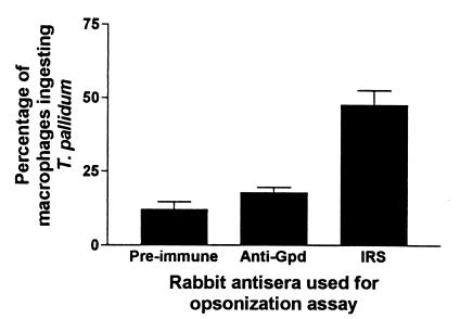
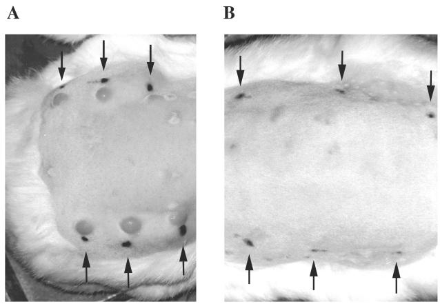
Infectious syphilis, caused by the spirochete bacterium Treponema pallidum subsp. pallidum, remains a public health concern worldwide. The immune-response evasion mechanisms employed by T. pallidum are poorly understood, and prior attempts to identify immunoprotective antigens for subsequent vaccine design have been unsuccessful. Previous investigations conducted in our laboratory identified the T. pallidum glycerophosphodiester phosphodiesterase as a potential immunoprotective antigen by using a differential immunologic expression library screen. In studies reported here, heterologous expression of the T. pallidum glycerophosphodiester phosphodiesterase in Escherichia coli yielded a full-length, enzymatically active protein. Characterization of the recombinant molecule showed it to be bifunctional, in that it exhibited specific binding to human immunoglobulin A (IgA), IgD, and IgG in addition to possessing enzymatic activity. IgG fractionation studies revealed specific binding of the recombinant enzyme to the Fc fragment of human IgG, a characteristic that may play a role in enabling the syphilis spirochete to evade the host immune response. In further investigations, immunization with the recombinant enzyme significantly protected rabbits from subsequent T. pallidum challenge, altering lesion development at the sites of challenge. In all cases, animals immunized with the recombinant molecule developed atypical pale, flat, slightly indurated, and nonulcerative reactions at the challenge sites that resolved before lesions appeared in the control animals. Although protection in the immunized rabbits was incomplete, as demonstrated by the presence of T. pallidum in the rabbit infectivity test, glycerophosphodiester phosphodiesterase nevertheless represents a significantly immunoprotective T. pallidum antigen and thus may be useful for inclusion in an antigen cocktail vaccine for syphilis.


Figures

FIG. 1
FIG. 1
Overexpression of the recombinant T. pallidum Gpd and analysis of anti-Gpd immunoreactivity. (A) Coomassie blue-stained SDS-PAGE analysis of E. coli BL21 (DE3) pLysS expressing either the Gpd-pET-3a construct (lane 1, crude lysate; lane 2, soluble fraction; lane 3, insoluble fraction) or the pET-3a vector alone (lane 4, insoluble fraction). (B) Immunoblot analysis of anti-Gpd immunoreactivity on purified inclusion bodies from E. coli BL21 (DE3) pLysS expressing the Gpd-pET-3a construct. Lanes: 1, anti-Gpd polyclonal antiserum, 2, E. coli-adsorbed anti-Gpd polyclonal antiserum. Each lane contains approximately 2 μg of total bacterial lysate and soluble or insoluble bacterial fractions, and molecular mass standards in kilodaltons are indicated at the left of each panel. In each panel, the recombinant T. pallidum Gpd is indicated by an arrow.
FIG. 2
FIG. 2
Opsonic potential of the recombinant T. pallidum Gpd. Shown are the percentages of rabbit peritoneal macrophages phagocytosing T. pallidum after a 4-h incubation with a 1:100 dilution of either IRS, anti-T. pallidum Gpd polyclonal antiserum, or preimmune serum (normal rabbit serum) collected prior to commencement of the immunization protocol. Three separate experiments were performed to test for opsonization with a total number of replicate assays of eight (preimmune serum), seven (anti-Gpd serum) and nine (IRS). Bars show standard errors of the means.
FIG. 3
FIG. 3
Immunoblot analysis of anti-Gpd immunoreactivity on T. pallidum lysates. Antiserum raised against the recombinant T. pallidum Gpd was used to probe an unwashed T. pallidum lysate (lane 1) and lysates of T. pallidum washed one (lane 2) and three (lane 3) times. Each lane contains 1.4 × 10⁷ treponemes, and molecular mass standards in kilodaltons are indicated at the left. The T. pallidum Gpd is indicated by an arrow. The strongly immunoreactive bands at approximately 55 and 25 kDa represent the heavy and light chains, respectively, of rabbit antibody molecules that are contaminating the T. pallidum lysate preparations and are detected by the goat F(ab′)2 anti-rabbit IgG secondary antibody. This immunoreactivity also decreases in washed treponemes.
FIG. 4
FIG. 4
Immunoblot analyses of the Ig-binding capability of recombinant T. pallidum Gpd. Shown are inclusion bodies purified from E. coli BL21 (DE3) pLysS transformed with either the pET-3a-Gpd construct (lanes 1, 3, 5, and 7) or with the pET-3a vector alone (lanes 2, 4, 6, and 8). Molecular mass standards in kilodaltons are indicated on the left of each panel. (A) Specificity of recombinant T. pallidum Gpd for binding various Ig classes. Lanes: 1 and 2, human IgA and goat F(ab′)2 anti-human IgA (alpha chain specific); 3 and 4, human IgD and goat F(ab′)2 anti-human IgD (delta chain specific); 5 and 6, human IgG and goat F(ab′)2 anti-human IgG (gamma chain specific); 7 and 8, human IgM and goat F(ab′)2 anti-human IgM (mu chain specific). (B) Investigation of the binding specificity of recombinant T. pallidum Gpd for human IgG. In all cases the secondary antibody used was goat F(ab′)2 anti-human IgG (gamma chain specific). Lanes: 1 and 2, intact human IgG; 3 and 4, human IgG Fc fragment; 5 and 6, human IgG Fab fragment; 7 and 8, no primary Ig.
FIG. 5
FIG. 5
Immunoprotective capacity of recombinant T. pallidum Gpd. Shown are representative rabbits from the control pET-3a immunization group (A) and the Gpd-pET-3a immunization group (B). The lesions observed on the unimmunized control rabbits were similar to those of the control pET-3a immunization group, and thus are not shown. The black ink spots indicated by arrows are adjacent to the intradermal challenge sites. The faint scars present on the back of the rabbits are sites of subcutaneous immunizations. Photographs were taken prior to lesion ulceration in the control rabbits on day 23 postchallenge.

Similar articles

Cited by

References

    1. Akins D R, Robinson E, Shevchenko D, Elkins C, Cox D L, Radolf J D. Tromp1, a putative rare outer membrane protein, is anchored by an uncleaved signal sequence to the Treponema pallidum cytoplasmic membrane. J Bacteriol. 1997;179:5076–5086. - PMC - PubMed
    1. Akkoyunlu M, Ruan M, Forsgren A. Distribution of protein D, an immunoglobulin D-binding protein, in Haemophilus strains. Infect Immun. 1991;59:1231–1238. - PMC - PubMed
    1. Alderete J F, Baseman J B. Surface-associated host proteins on virulent Treponema pallidum. Infect Immun. 1979;26:1048–1056. - PMC - PubMed
    1. Baker-Zander S A, Hook E W, Bonin P, Handsfield H H, Lukehart S A. Antigens of Treponema pallidum recognized by IgG and IgM antibodies during syphilis infection in humans. J Infect Dis. 1985;151:264–272. - PubMed
    1. Baker-Zander S A, Lukehart S A. Macrophage-mediated killing of opsonized Treponema pallidum. J Infect Dis. 1992;165:69–74. - PubMed

Publication types

MeSH terms