

Intranasal immunization with cytotoxic T-lymphocyte epitope peptide and mucosal adjuvant cholera toxin: selective augmentation of peptide-presenting dendritic cells in nasal mucosa-associated lymphoid tissue
- PMID: 9826368
- PMCID: PMC108744
- DOI: 10.1128/IAI.66.12.5876-5881.1998
Intranasal immunization with cytotoxic T-lymphocyte epitope peptide and mucosal adjuvant cholera toxin: selective augmentation of peptide-presenting dendritic cells in nasal mucosa-associated lymphoid tissue
Abstract
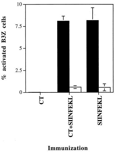
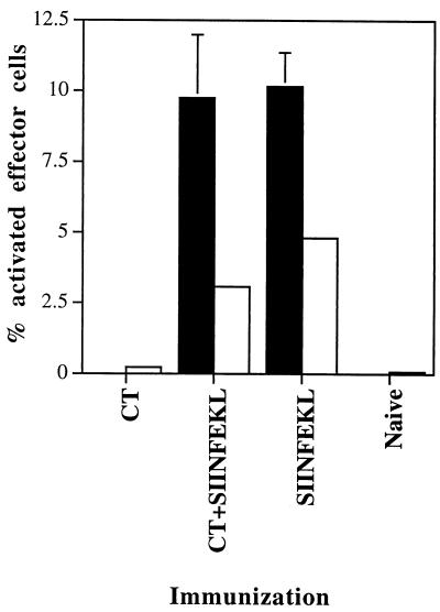
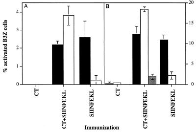
We previously reported that cholera toxin (CT) was required as a mucosal adjuvant for the induction of peptide-specific cytotoxic T lymphocytes (CTL) following intranasal immunization with CTL epitope peptides (A. Porgador et al., J. Immunol. 158:834-841, 1997). The present study was performed to identify the site and the antigen-presenting cell (APC) population responsible for the presentation of intranasally administered CTL epitope peptide immunogens and to determine whether CT directly affects antigen presentation by these APCs. For these experiments, C57BL/6 mice were intranasally immunized with the ovalbumin H-2Kb-restricted CTL epitope SIINFEKL with or without CT. Cells were then isolated from the cervical lymph nodes (CLN) and the nasal mucosa-associated lymphoid tissue (NALT) and tested for the ability to stimulate the B3Z T-cell hybridoma, which recognizes SIINFEKL in association with H-2Kb. Dendritic cell (DC)-enriched CLN cells from mice immunized with peptide and CT or peptide only could stimulate B3Z cells, while DC-depleted CLN cells from either group were unable to stimulate B3Z cells. NALT cells of mice immunized with peptide and CT, but not with peptide alone, were able to efficiently stimulate B3Z hybridomas. Depletion of N418-positive DC from these NALT cells resulted in significant reduction of B3Z activation. Our results indicate that DC are the APC responsible for the presentation of CTL epitope peptides following intranasal immunization and that CT augments the ability of dendritic cells in the NALT, but not in the draining CLN, to present CLT epitope peptides. This finding suggests that CT acts locally as a mucosal adjuvant and that NALT DC are the predominant APC involved with the induction of immunity after intranasal immunization with peptide immunogens and CT.
Figures
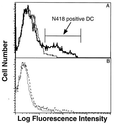
 , DC-depleted NALT.
References
-
- Bromander A, Holmgren J, Lycke N. Cholera toxin stimulates IL-1 production and enhances antigen presentation by macrophages in vitro. J Immunol. 1991;146:2908–2914. - PubMed
-
- Bromander A K, Kjerrulf M, Holmgren J, Lycke N. Cholera toxin enhances alloantigen presentation by cultured intestinal epithelial cells. Scand J Immunol. 1993;37:452–458. - PubMed
-
- Chen Y, Inobe J, Marks R, Gonnella P, Kuchroo V K, Weiner H L. Peripheral deletion of antigen-reactive T cells in oral tolerance. Nature. 1995;376:177–180. . (Erratum, 21:377.) - PubMed
MeSH terms
Substances
LinkOut - more resources
Full Text Sources
Other Literature Sources
Miscellaneous
