. 1998 Dec;66(12):5906-14.
doi: 10.1128/IAI.66.12.5906-5914.1998.

Streptococcus sanguis-induced platelet clotting in rabbits and hemodynamic and cardiopulmonary consequences

Affiliations

Streptococcus sanguis-induced platelet clotting in rabbits and hemodynamic and cardiopulmonary consequences

M W Meyer et al. Infect Immun. 1998 Dec.

Abstract

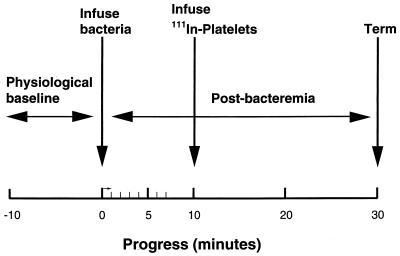
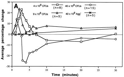
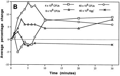
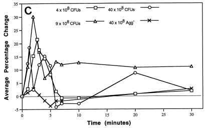
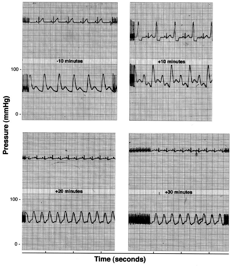
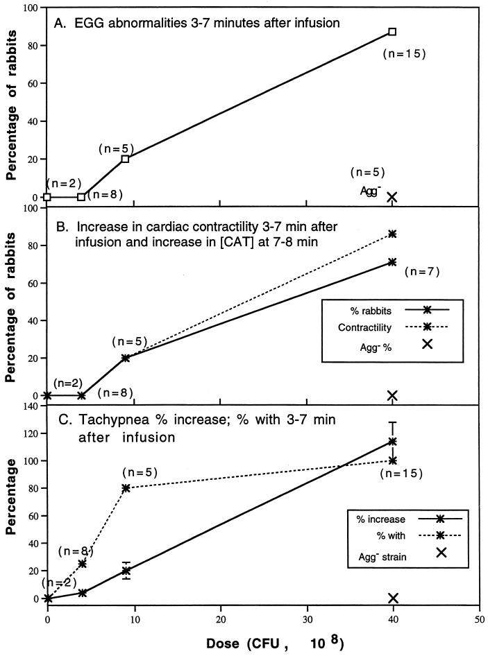
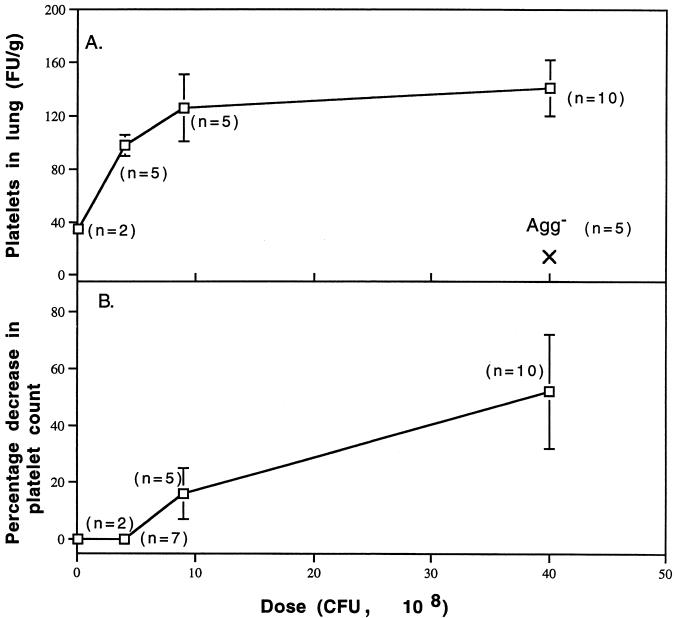
By mimicking hemostatic structural domains of collagen, Streptococcus sanguis (aggregation-positive phenotype; Agg+) induces platelets to aggregate in vitro. To test the hypothesis that aggregation occurs in vivo, S. sanguis (Agg+ or Agg- suspension) was infused intravenously into rabbits. The extent of hemodynamic and cardiopulmonary changes and the fate of circulating platelets were Agg+ strain dose dependent. Within 45 to 50 s of the start of infusion, 40 x 10(8) CFU of the Agg+ strain caused increased blood pressure. Thirty seconds after infusion, other changes occurred. Intermittent electrocardiographic abnormalities (13 of 15 rabbits), ST-segment depression (10 of 15 rabbits), and preventricular contractions (7 of 15 rabbits) manifested at 3 to 7 min, with frequencies dose dependent. Respiratory rate and cardiac contractility increased during this phase. Blood catecholamine concentration, thrombocytopenia, accumulation of 111Indium-labeled platelets in the lungs, and ventricular axis deviation also showed dose dependency. Rabbits were unaffected by inoculation of an Agg- strain. Therefore, Agg+ S. sanguis induced platelet aggregation in vitro. Platelet clots caused hemodynamic changes, acute pulmonary hypertension, and cardiac abnormalities, including ischemia.


Figures

FIG. 1
FIG. 1
Experimental protocol. To study the consequences of S. sanguis infusion in rabbits, physiological variables were measured or monitored for pre- (10 min) and postbacteremia (30 min) periods. Ten minutes after induction of the bacteremia, 111Indium-labeled donor platelets were infused to assess circulating platelets. Figure reproduced with modifications (31) with permission of the publisher.
FIG. 2
FIG. 2
Hemodynamic consequences of S. sanguis bacteremia. The hemodynamic status over time was monitored. Blood pressure and heart rate were normalized as percentage changes over time as a function of dose of S. sanguis. The heart rate × blood pressure product (an index of cardiac energy and hemodynamic status) was normalized and plotted. The number of rabbits (n) used to establish the mean for each dose is indicated in panel A. For each dose, significant differences from the baseline were determined by the Student t test. (A) Arterial blood pressure. Average percentage change with the Agg+ strain was plotted for each minute of the first 7 min, and at 10, 20, and 30 min. The time and value of the maximal increase was plotted for each dose. Changes ≥± 8% were significantly different from baseline or zero. All values calculated for the Agg strain were not different from zero. (B) Heart rate. Average percentage change was plotted at the same minute intervals as blood pressure. Changes ≥± 4 to 5% were significantly different from zero. The Agg strain, even at 40 × 108 CFU, did not alter the heart rate during the period of bacteremia. (C) Heart rate × blood pressure. The average percentage change was plotted over time as above. Changes ≥± 8% were significantly different from zero. Panels A and B have been reproduced with modifications (30) with permission of the publisher.
FIG. 2
FIG. 2
Hemodynamic consequences of S. sanguis bacteremia. The hemodynamic status over time was monitored. Blood pressure and heart rate were normalized as percentage changes over time as a function of dose of S. sanguis. The heart rate × blood pressure product (an index of cardiac energy and hemodynamic status) was normalized and plotted. The number of rabbits (n) used to establish the mean for each dose is indicated in panel A. For each dose, significant differences from the baseline were determined by the Student t test. (A) Arterial blood pressure. Average percentage change with the Agg+ strain was plotted for each minute of the first 7 min, and at 10, 20, and 30 min. The time and value of the maximal increase was plotted for each dose. Changes ≥± 8% were significantly different from baseline or zero. All values calculated for the Agg strain were not different from zero. (B) Heart rate. Average percentage change was plotted at the same minute intervals as blood pressure. Changes ≥± 4 to 5% were significantly different from zero. The Agg strain, even at 40 × 108 CFU, did not alter the heart rate during the period of bacteremia. (C) Heart rate × blood pressure. The average percentage change was plotted over time as above. Changes ≥± 8% were significantly different from zero. Panels A and B have been reproduced with modifications (30) with permission of the publisher.
FIG. 3
FIG. 3
Representative electrocardiographic tracing. Selected segments of an ECG and blood pressure recording for a rabbit given 40 × 108 CFU of the Agg+ strain is shown. Note ST-segment depression and alternating preventricular contractions (PVCs) by 10 min. The ST-segment depression persists through t = 30 min, the heart rate remains elevated, and the pulse pressure is greatly reduced.
FIG. 4
FIG. 4
Cardiopulmonary consequences of S. sanguis bacteremia. For selected variables, the Agg+ dose-dependent changes in frequency or magnitude are presented. Two rabbits infused with 0.1 × 10⁸ CFU of the Agg+ strain and five with 40 × 108 CFU of the Agg strain are also included for comparison. Note that the Agg strain had no effect upon any of the cardiopulmonary variables studied. The regression line for each variable shown was significantly different from zero (linear regression analysis). (A) ECG abnormalities at t = 3 to 7 min. During this period after infusion, the ECG was monitored and changes were evaluated for the numbers of rabbits shown. (B) Increases in cardiac contractility (t = 3 to 7 min) and catacholamine concentration (t = 7 to 8 min). Cardiac contractility was computed as dP/dt as described in Materials and Methods. In the same rabbits, catacholamine concentrations were determined by radioenzymatic assay as described in Materials and Methods. (C) Frequency and percent increase in tachypnea at t = 3 to 7 min. Note the high frequency but comparatively low magnitude with an Agg+ dose of 9 × 10⁸ CFU. Catecholamines did not vary from baseline in rabbits given Agg+ doses of 0.1 and 4 × 10⁸ CFU. Since the cardiopulmonary responses were similar to rabbits given lower doses of the Agg+ strain, rabbits given 40 × 108 CFU of the Agg strain were not sampled for catecholamines.
FIG. 5
FIG. 5
Platelet fate. Dose-dependent signs of platelet aggregation in vivo were quantified. (A) Fractional uptake (FU/g) of 111Indium-labeled platelets in the lungs at t = 30 min. The FU/gram of lung becomes saturated at doses larger than 9 × 10⁸ CFU of the Agg+ strain (no difference between doses; Student’s t test). In those rabbits given 40 × 108 CFU of the Agg strain, the FU/gram of lung was nearly three-fold less than in the two rabbits given 0.1 × 10⁸ CFU of the Agg+ strain. (B) Percentage decrease in platelet count. Platelets were counted at baseline and at t = 30 min as described in Materials and Methods.

Similar articles

Cited by

References

    1. Adler D, Nikolic S D, Sonnenbick E H, Yellin E L. Time to dP/dtmax, a preload-independent index of contractility: open-chest dog study. Basic Res Cardiol. 1996;91:94–100. - PubMed
    1. Angrist A A, Oka M, Nakao K. Vegetative endocarditis. Pathol Annu. 1967;2:155–212.
    1. Bancsi M J L M F, Thompson J, Bertina R M. Stimulation of monocyte tissue factor expression in an in vitro model of bacterial endocarditis. Infect Immun. 1994;62:5669–5672. - PMC - PubMed
    1. Bancsi M J, Veltrop M H, Bertina R M, Thompson J. Influence of monocytes and antibiotic treatment on tissue factor activity of endocardial vegetation in rabbits infected with Streptococcus sanguis. Infect Immun. 1996;64:448–451. - PMC - PubMed
    1. Beck, J., R. Garcia, G. Heiss, P. S. Vokonas, and S. Offenbacher. 1996. Periodontal disease and cardiovascular disease. J. Periodontol. 67(Suppl.; symposium proceedings):1123–1137. - PubMed

Publication types