. 1998 Dec;66(12):5948-54.
doi: 10.1128/IAI.66.12.5948-5954.1998.

Detection of anionic antimicrobial peptides in ovine bronchoalveolar lavage fluid and respiratory epithelium

Affiliations

Detection of anionic antimicrobial peptides in ovine bronchoalveolar lavage fluid and respiratory epithelium

K A Brogden et al. Infect Immun. 1998 Dec.

Abstract

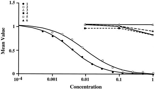
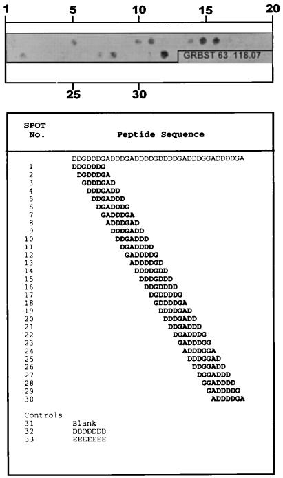
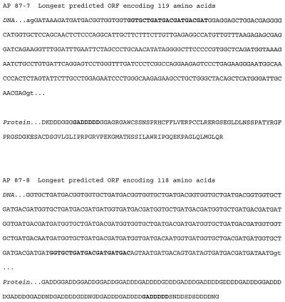
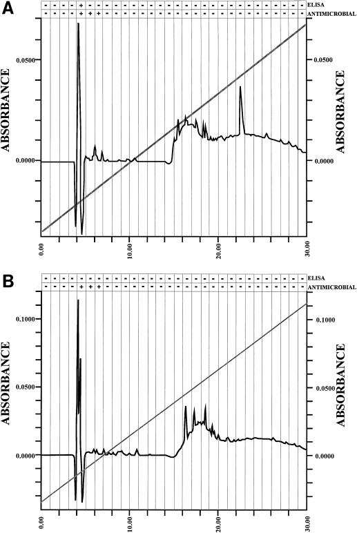
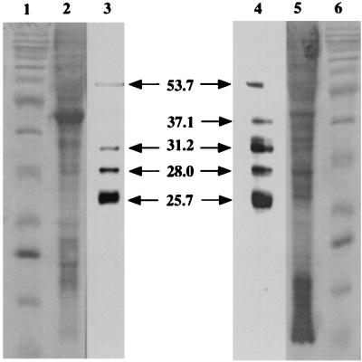
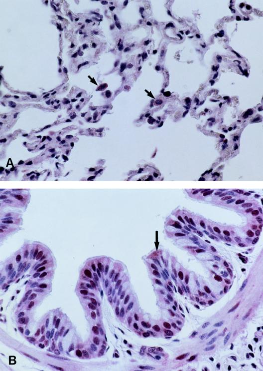
Three small antimicrobial anionic peptides (AP) were originally isolated from an ovine pulmonary surfactant. However, their presence in bronchoalveolar lavage (BAL) fluid and tissues of the respiratory tract is unknown. In this study, we made affinity-purified rabbit polyclonal and mouse monoclonal antibodies to synthetic H-DDDDDDD-OH. Antibody specificity was assessed by a competitive enzyme-linked immunosorbent assay (ELISA), and the exact epitope binding sites were determined with analog peptides synthesized on derivatized cellulose. These antibodies were used to detect AP in BAL fluid by ELISA and in respiratory tissues by Western blot analysis and immunocytochemistry. BAL fluid from 25 sheep contained 0.83 +/- 0.33 mM AP (mean +/- standard deviation; range, 0.10 to 1.59 mM) and was antimicrobial. The presence of AP in BAL fluid was confirmed by reverse-phase high-pressure liquid chromatography fractionation followed by matrix-assisted laser desorption/ionization (MALDI) mass spectrometry on those fractions which were positive by competitive ELISA and demonstrated antimicrobial activity. In Western blots, polyclonal antibody PAB96-1 and monoclonal antibody 1G9-1C2 (5.0 micrograms/ml) detected four bands in solubilized turbinate and tracheal epithelial cells (53.7, 31.2, 28.0, and 25.7 kDa) and five bands in lung homogenates (53.5, 37.1, 31.2, 28.0, and 25.7 kDa). Only a single band was seen in solubilized liver and small-intestine homogenates, and no bands were seen in blots containing BAL fluid, albumin, or kidney or spleen homogenates. In pulmonary-tissue sections, both antibodies PAB96-1 and 1G9-1C2 identified accumulated protein in the apical cytoplasm of the bronchial and bronchiolar epithelia, in the cytoplasm of pulmonary endothelial cells, and in an occasional alveolar macrophage. As a first step in identifying a candidate AP precursor gene(s), degenerate oligonucleotides representing all possible coding combinations for H-GADDDDD-OH and H-DDDDDDD-OH were synthesized and used to probe Southern blots of sheep genomic DNA. Following low-stringency washes and a 2-day exposure, strongly hybridizing bands could be identified. One degenerate oligonucleotide, SH87, was used as a hybridization probe to screen a sheep phage genomic library. Two independent phage contained the H-GADDDDD-OH coding sequence as part of a larger predicted protein. AP may originate as part of an intracellular precursor protein, with multistep processing leading to the release of the heptapeptide into mucosal secretions. There it may interact with other innate pulmonary defenses to prevent microbial infection.


Figures

FIG. 1
FIG. 1
The competitive ELISA was used to assess the specificity of monoclonal antibody 1G9-1C2. BSA-DDDDDDD-OH conjugate (50 ng of conjugate/well) was used as the adsorbed antigen, and varying graded concentrations (1.0 to 0.002 mM) of H-DDDDDDD-OH (peptide 1), H-GADDDDD-OH (peptide 2), H-TQDDGGK-OH (peptide 3), H-GGEEK-OH (peptide 4), H-VDDDDK-OH (peptide 5), and H-SGSGSGS-OH (peptide 6) were used as the competitive antigens in the presence of antibody 1G9-1C2 (1.0 μg/ml). Antibody 1G9-1C2 was specific and recognized H-GADDDDD-OH nearly as well as H-DDDDDDD-OH. However, the sequences with internal Asp or Glu residues (e.g., H-VDDDDK-OH, H-TQDDGGK-OH, and H-GGEEK-OH) were not recognized and did not cross-react. H-SGSGSGS-OH was a negative-control peptide.
FIG. 2
FIG. 2
The epitope binding sites of antibody 1G9-1C2 (25 μg/ml) were determined with 30 peptides (7 residues each) corresponding to a single-residue frameshift of the 7-residue sequence starting at residue 30 of the sequence identified in AP 87-8 of Fig. 3. For example, peptide 1 was H-DDGDDDG-OH, peptide 2 was H-DGDDDGA-OH, peptide 3 was H-GDDDGAD-OH, etc. Peptides were synthesized simultaneously on a derivatized cellulose sheet (SPOTs; Genosys). SPOT 32 contained H-DDDDDDD-OH and SPOT 33 contained H-EEEEEEE-OH as peptide controls. SPOTs were detected after the strip was incubated in casein-based blocking buffer, incubated in antibody, washed, incubated in secondary β-galactosidase-conjugated antibody, and incubated in Signal Development solution. Epitopes that contained more than 2 terminal Asp residues were recognized.
FIG. 3
FIG. 3
Nucleotide and predicted peptide sequences for candidate AP precursor genes, comprising exons from sheep genomic library clones. The predicted ORF for AP 87-8 extends in the 5′ direction. ORF sequences are capitalized, and potential AP coding sequences are in boldface.
FIG. 4
FIG. 4
RP-HPLC chromatograms of ovine BAL fluid (A) and ovine epithelial-cell extracts (B), showing peak fraction 5, eluted in 13 to 14% acetonitrile, with antimicrobial activity and evidence of AP by competitive ELISA with antibody 1G9-1C2. BSA-DDDDDDD-OH conjugate (50 ng of conjugate/well) was used as the adsorbed antigen, and RP-HPLC fractions were used as the competitive antigens in the presence of antibody 1G9-1C2 (1.0 μg/ml). To confirm the presence of AP, fractions were examined by MALDI mass spectrometry. H-DDDDDDD-OH [(M + H)+; 824.4 Da], H-GDDDDDD-OH [(M + H)+; 766.1 Da], and H-GADDDDD-OH [(M + H)+; 718.24 Da] were detected.
FIG. 5
FIG. 5
Western blot of ovine tracheal epithelium and lung homogenate. Lane 1, protein ladder (10 to 200 kDa) standard; lane 2, tracheal-cell homogenate stained with Coomassie blue; lane 3, tracheal-cell homogenate probed with antibody PAB96-1; lane 4, lung homogenate probed with antibody PAB96-1; lane 5, lung homogenate stained with Coomassie blue; lane 6, protein ladder standard. In tracheal epithelial cells, antibody PAB96-1 identified four bands with molecular masses of 53.7, 31.2, 28.0, and 25.7 kDa. In the lung, antibody PAB96-1 identified five bands with molecular masses of 53.5, 37.1, 31.2, 28.0, and 25.7 kDa. Identical results were seen with antibody 1G9-1C2 (data not shown). As controls, strips of membrane containing separated tracheal and lung proteins were incubated with preimmunized rabbit serum (1:200), preimmunized mouse serum (1:200), or peroxidase-labeled goat anti-rabbit IgG (0.5 μg/ml) or goat anti-mouse IgG (0.5 μg/ml). No reactions were seen (data not shown). In addition, preincubation of antibody PAB96-1 (5 μg/ml) or 1G9-1C2 (5 μg/ml) with 1.0 mM H-DDDDDDD-OH eliminated specific staining of all bands (data not shown). Blots were photographed (digital camera RD-175; Minolta), and the sizes of the reactive bands were determined (GelCompar, v. 4.0; Applied Maths) by extrapolation from a standard curve of the sizes of the stained bands on the protein ladder.
FIG. 6
FIG. 6
Tissue sections of ovine lung, incubated with antibody 1G9-1C2, show immunocytochemically stained regions of the alveoli (A) and bronchial epithelium (B) that were not seen in these regions when tissue sections were incubated in secondary antiserum alone (data not shown). Antibody 1G9-1C2 identified accumulated protein (arrows) in the cytoplasm of pulmonary endothelial cells and an occasional alveolar macrophage (A) and in the apical cytoplasm of the bronchial and bronchiolar epithelia (B). Goblet cells and epithelial cells of pulmonary alveoli were not stained.

References

    1. Bartlett J G. Bacteriological diagnosis of pulmonary infections. In: Sackner M A, editor. Diagnostic techniques in pulmonary disease. Vol. 16. New York, N.Y: Marcel Dekker, Inc.; 1981. pp. 707–745.
    1. Bligh E G, Dyer W J. A rapid method of total lipid extraction and purification. Can J Biochem Physiol. 1959;37:911–917. - PubMed
    1. Boman H G. Peptide antibiotics and their role in innate immunity. Annu Rev Immunol. 1995;13:61–92. - PubMed
    1. Brogden K A, Ackermann M, Huttner K M. Small, anionic, and charge-neutralizing propeptide fragments of zymogens are antimicrobial. Antimicrob Agents Chemother. 1997;41:1615–1617. - PMC - PubMed
    1. Brogden K A, Ackermann M R, DeBey B M. Pasteurella haemolytica lipopolysaccharide-associated protein induces pulmonary inflammation after bronchoscopic deposition in calves and sheep. Infect Immun. 1995;63:3595–3599. - PMC - PubMed

MeSH terms