. 1998 Dec;66(12):5972-9.
doi: 10.1128/IAI.66.12.5972-5979.1998.

Evidence for multiple pathologic and protective mechanisms of murine cerebral malaria

Affiliations

Evidence for multiple pathologic and protective mechanisms of murine cerebral malaria

V M Jennings et al. Infect Immun. 1998 Dec.

Abstract

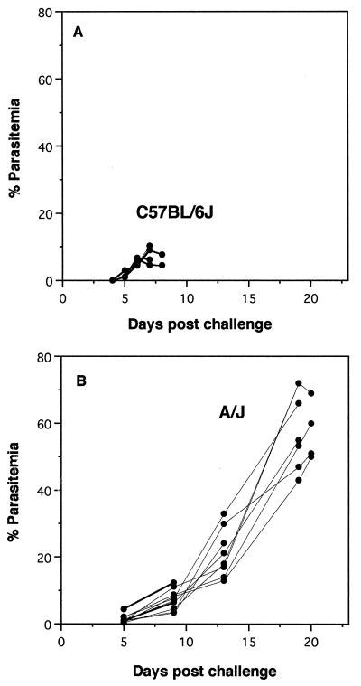
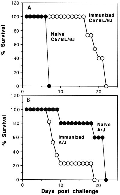
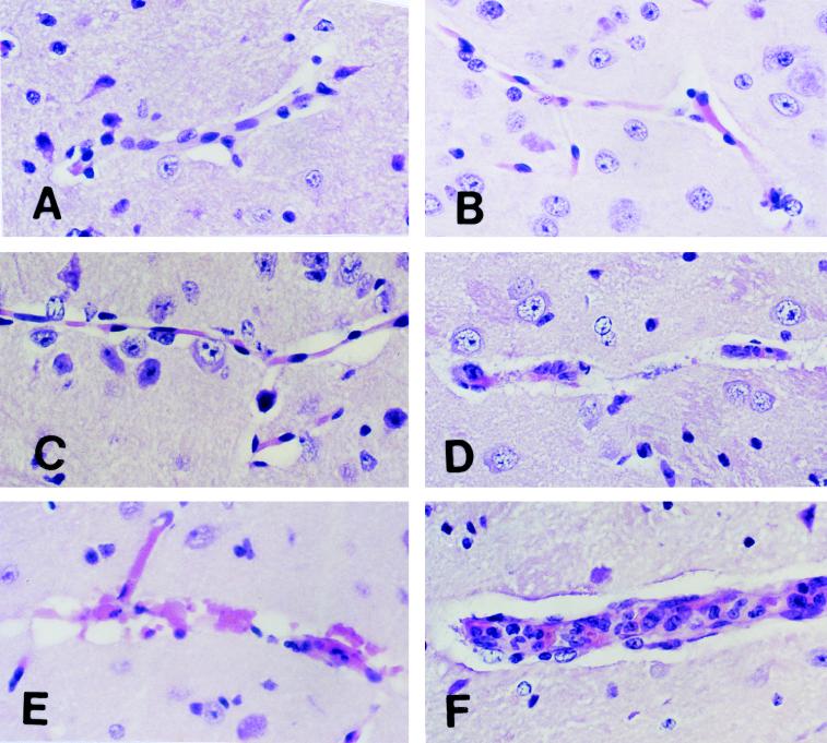
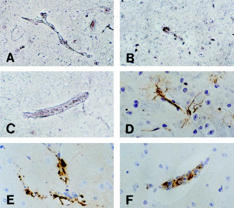
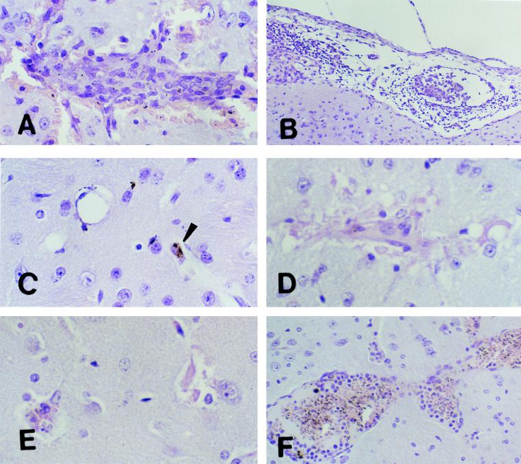
Murine cerebral malaria (CM) induced by Plasmodium berghei ANKA kills susceptible mice within 24 to 48 h of onset of symptoms and is characterized by the production of inflammatory cytokines in the brain. C57BL/6J mice are sensitive to lethal CM, while A/J mice are resistant. These strains of mice were immunized with an adjuvant vaccine of killed whole-blood-stage parasites. The immunization protected C57BL/6 mice from lethal CM following virulent challenge. The same immunization increased the incidence of lethal CM in A/J mice challenged similarly. Histopathologic examination of the brains of mice from these studies revealed two distinct types of lesions. Type I CM is acute in onset; usually lethal; and characterized by widespread microglial activation, endothelial cell damage, and microvascular disruption in the brain. Type II CM is characterized by intense, but focal, mononuclear cell inflammation without endothelial cell damage or microvascular destruction. Animals with type II lesions were clinically normal and protected from type I lesions. Available clinical, epidemiological, and biochemical evidence suggests that type I and type II lesions might exist in human CM as well.


Figures

FIG. 1
FIG. 1
Parasitemia of unimmunized infected mice. Groups of C57BL/6J and A/J mice were challenged intravenously with 104 P. berghei ANKA-parasitized erythrocytes. Percentages of erythrocytes with parasites were determined by examination of thin blood smears from individual mice taken at 2- to 4-day intervals until death. The groups are C57BL/6J mice (n = 10), which are susceptible to CM (A), and A/J mice (n = 10), which are resistant to CM (B).
FIG. 2
FIG. 2
Effect of immunization on survival following infection. Groups of C57BL/6J and A/J mice were immunized three times subcutaneously with a whole-blood-stage P. berghei vaccine. Percentages of survival of naive control (filled circles) and immunized (open circles) groups of C57BL/6J (n = 15) (A) and A/J (n = 17) (B) mice following challenge infection with 104 parasitized erythrocytes are shown.
FIG. 3
FIG. 3
Morphology of CM lesions in the brains of C57BL/6J mice. Normal C57BL/6J mice were challenged intravenously with 104 parasitized erythrocytes. Brains were removed for histologic examination at different times. (A) Section of normal mouse brain showing a healthy unaffected blood vessel. (B) Section of mouse brain on day 3 after challenge showing microglial cells in the perivascular space. (C) Section of mouse brain on day 5 after challenge showing increased numbers of activated microglial cells in perivascular spaces. (D and E) Sections of mouse brain on day 7 after challenge showing destruction of endothelial cells and disruption of vessel walls. Endothelial and other cell nuclei were occasionally condensed and/or fragmented in a fashion consistent with apoptosis. (F) Section of mouse brain on day 7 after challenge showing mononuclear cell accumulation and endothelial cell activation in a blood vessel from the brain of a mouse moribund with CM. These were focal lesions. Larger areas of the brains of the same mice demonstrated the changes shown in panels D and E. All sections were stained with H&E. Magnification, ×500.
FIG. 4
FIG. 4
Reticulum and HAM-56 stainings of CM lesions in the brains of C57BL/6J mice. (A) Section of normal mouse brain showing a healthy uninterrupted blood vessel reticulum. (B) Section of mouse brain on day 7 after challenge showing destruction of reticulum fibers of a vessel. This change was characteristic of vessels with the lesions shown in Fig. 3D and E. (C) Section of mouse brain on day 7 after challenge showing preservation of reticulum fibers around a blood vessel with monocyte inflammation. This pattern of reticulum staining was characteristic of vessels with the lesions shown in Fig. 3F. Magnification, ×200. (D) Section of normal mouse brain showing a HAM-56 monoclonal antibody staining of endothelial cells and glial processes. (E) Section of mouse brain on day 7 after challenge showing a HAM-56 staining of aggregates of material associated with disrupted blood vessels. This pattern of staining was characteristic of vessels with the lesions shown in Fig. 3D and E. (F) Section of mouse brain on day 7 after challenge showing a HAM-56 staining of monocytes in an intact blood vessel with monocyte inflammation. This pattern of staining was characteristic of vessels with the lesions shown in Fig. 3F. All sections were stained with H&E. Magnification, ×200.
FIG. 5
FIG. 5
Morphologies of lesions in the brains of immunized C57BL/6J (A to C) and A/J (D to F) mice. Immunized C57BL/6J and A/J mice were challenged intravenously with 104 parasitized erythrocytes. C57BL/6J mice were clinically normal, with no signs of CM. In contrast, some A/J mice developed CM while others remained clinically normal, with no signs of CM. Brains were removed from both groups for histologic examination at different times. (A) Section of mouse brain on day 10 after challenge showing an inflammatory hemorrhagic vascular lesion in the cortex. Magnification, ×500. (B) Section of mouse brain and meninges on day 10 after challenge showing meningitis. The monocyte was the predominant inflammatory cell within both vessels and surrounding tissue. Magnification, ×200. (C) Section of mouse brain 21 days after challenge showing resolution and reversibility of the inflammatory and hemorrhagic lesions. Small hemosiderin deposits (arrowhead) are the only remnants of previous lesions. Magnification, ×500. (D and E) Sections of the brains of mice moribund with CM on day 8 after challenge. Changes in microglial cells, destruction of endothelial cells, and disruption of the vessel walls similar to that observed in C57BL/6J mice with CM are evident. The sections appear pale because of loss of cellular integrity. Magnification, ×500. (F) Section of the brain of a mouse protected from lethal CM on day 18 after challenge showing an intense inflammatory infiltrate within blood vessels made up primarily of activated lymphocytes, macrophages, and malarial pigment but in which the endothelium remains intact. These were focal lesions. Areas of the brains of these mice away from these lesions appeared normal. Magnification, ×200. All sections were stained with H&E.

References

    1. Adams C W, Poston R N. Macrophage histology in paraffin-embedded multiple sclerosis plaques is demonstrated by the monoclonal panmacrophage marker HAM-56: correlation with chronicity of the lesion. Acta Neuropathol. 1990;80:208–211. - PubMed
    1. Allen S, Genton B, Alexander N, Gibson N, Raiko A. Malaria vaccine in children under 12 months of age. Lancet. 1995;346:1555–1556. - PubMed
    1. Barnwell J W, Howard R J, Coon H G, Miller L H. Splenic requirement for antigenic variation and expression of the variant antigen on the erythrocyte membrane in cloned Plasmodium knowlesi malaria. Infect Immun. 1983;40:985–994. - PMC - PubMed
    1. Clark I A, Ilschner S, MacMicking J D, Cowden W B. TNF and Plasmodium berghei ANKA-induced cerebral malaria. Immunol Lett. 1990;25:195–198. - PubMed
    1. Curfs J H, Hermsen C C, Meuwissen J H, Eling W M. Immunization against cerebral pathology in Plasmodium berghei-infected mice. Parasitology. 1992;105:7–14. - PubMed

Publication types

Substances

LinkOut - more resources