

Selective activation of sar promoters with the use of green fluorescent protein transcriptional fusions as the detection system in the rabbit endocarditis model
- PMID: 9826382
- PMCID: PMC108758
- DOI: 10.1128/IAI.66.12.5988-5993.1998
Selective activation of sar promoters with the use of green fluorescent protein transcriptional fusions as the detection system in the rabbit endocarditis model
Abstract
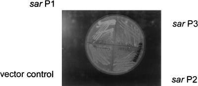
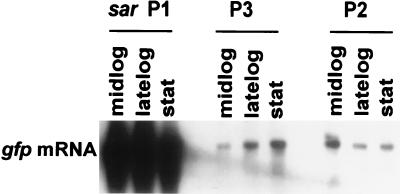
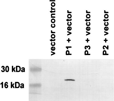
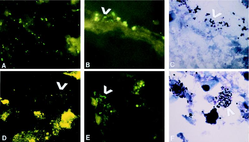
The global regulatory locus sar is composed of three overlapping transcripts initiated from a triple-promoter system (designated P1, P3, and P2). To explore if the individual sar promoters are differentially expressed in vitro and in vivo, we constructed a shuttle plasmid (pALC1434) containing a promoterless gfpUV gene (a gfp derivative [Clontech]) preceded by a polylinker region. Recombinant shuttle vectors containing individual sar promoters upstream of the gfpUV reporter gene were then introduced into Staphylococcus aureus RN6390. Northern and immunoblot analysis revealed that P1 is stronger than the P2 and P3 promoters in vitro. Additionally, the levels of the gfpUV transcript driven by individual sar promoters also correlated with the growth cycle dependency of these promoters in liquid cultures, thus suggesting the utility of pALC1434 as a vehicle for reporter fusion. Using the rabbit endocarditis model, we examined the expression of these three GFPUV fusions in vivo by fluorescence microscopy of infected cardiac vegetations 24 h after initial intravenous challenge. Similar to the in vitro findings, P1 was activated both in the center and on the surface of the vegetations. In contrast, the P3 promoter was silent both in vivo and in vitro as determined by fluorescence microscopy. Remarkably, P2 was silent in vitro but became highly activated in vivo. In particular, the sar P2 promoter was activated on the surface of the vegetation but not in the center of the lesion. These data imply that in vivo promoter activation of sar differed from that observed in vitro. Moreover, the individual sar promoters may be differentially expressed in different areas within the same anatomic niche, presumably reflecting the microbial physiological response to distinct host microenvironments. As the sar locus controls the synthesis of both extracellular and cell wall virulence determinants, these promoter-gfpUV constructs should be useful to characterize many aspects of S. aureus gene regulation in vivo.
Figures
Similar articles
-
Transcriptional analysis of different promoters in the sar locus in Staphylococcus aureus.J Bacteriol. 1998 Aug;180(15):3828-36. doi: 10.1128/JB.180.15.3828-3836.1998. J Bacteriol. 1998. PMID: 9683479 Free PMC article.
-
Characterization of sarR, a modulator of sar expression in Staphylococcus aureus.Infect Immun. 2001 Feb;69(2):885-96. doi: 10.1128/IAI.69.2.885-896.2001. Infect Immun. 2001. PMID: 11159982 Free PMC article.
-
The molecular architecture of the sar locus in Staphylococcus aureus.J Bacteriol. 1996 Aug;178(15):4563-70. doi: 10.1128/jb.178.15.4563-4570.1996. J Bacteriol. 1996. PMID: 8755885 Free PMC article.
-
Molecular analysis and organization of the sigmaB operon in Staphylococcus aureus.J Bacteriol. 2005 Dec;187(23):8006-19. doi: 10.1128/JB.187.23.8006-8019.2005. J Bacteriol. 2005. PMID: 16291674 Free PMC article.
-
Regulation of Staphylococcus aureus type 5 capsular polysaccharides by agr and sarA in vitro and in an experimental endocarditis model.Microb Pathog. 2002 Aug;33(2):73-9. doi: 10.1006/mpat.2002.0513. Microb Pathog. 2002. PMID: 12202106
Cited by
-
Influence of a functional sigB operon on the global regulators sar and agr in Staphylococcus aureus.J Bacteriol. 2001 Sep;183(17):5171-9. doi: 10.1128/JB.183.17.5171-5179.2001. J Bacteriol. 2001. PMID: 11489871 Free PMC article.
-
Following pathogen development and gene expression in a food ecosystem: the case of a Staphylococcus aureus isolate in cheese.Appl Environ Microbiol. 2014 Aug;80(16):5106-15. doi: 10.1128/AEM.01042-14. Epub 2014 Jun 13. Appl Environ Microbiol. 2014. PMID: 24928871 Free PMC article.
-
Persister formation in Staphylococcus aureus is associated with ATP depletion.Nat Microbiol. 2016;1:16051. doi: 10.1038/nmicrobiol.2016.51. Epub 2016 Apr 18. Nat Microbiol. 2016. PMID: 27398229 Free PMC article.
-
Identification and Application of a Panel of Constitutive Promoters for Gene Overexpression in Staphylococcus aureus.Front Microbiol. 2022 Feb 28;13:818307. doi: 10.3389/fmicb.2022.818307. eCollection 2022. Front Microbiol. 2022. PMID: 35295303 Free PMC article.
-
Role of sigmaB in the expression of Staphylococcus aureus cell wall adhesins ClfA and FnbA and contribution to infectivity in a rat model of experimental endocarditis.Infect Immun. 2005 Feb;73(2):990-8. doi: 10.1128/IAI.73.2.990-998.2005. Infect Immun. 2005. PMID: 15664942 Free PMC article.
References
-
- Blake M S, Johnston K H, Russell-Jones G J, Gotschlich E C. A rapid sensitive method for detection of alkaline phosphatase-conjugated anti-antibody on Western blots. Anal Biochem. 1984;136:175–179. - PubMed
-
- Boyce J M. Epidemiology and prevention of nosocomial infections. In: Crossley K B, Archer G L, editors. The staphylococci in human disease. New York, N.Y: Churchill Livingstone; 1997. pp. 309–329.
-
- Bradford M M. A rapid and sensitive method for the quantitation of microgram quantities of protein utilizing the principle of protein-dye binding. Anal Biochem. 1976;72:248. - PubMed
Publication types
MeSH terms
Substances
Grants and funding
LinkOut - more resources
Full Text Sources
Other Literature Sources
Medical
Research Materials
Miscellaneous
