

Comparative Study
doi: 10.1128/IAI.66.12.6017-6021.1998.
Chlamydia trachomatis IncA is localized to the inclusion membrane and is recognized by antisera from infected humans and primates
Affiliations
- PMID: 9826388
- PMCID: PMC108764
- DOI: 10.1128/IAI.66.12.6017-6021.1998
Item in Clipboard
Comparative Study
Chlamydia trachomatis IncA is localized to the inclusion membrane and is recognized by antisera from infected humans and primates
Infect Immun.
1998 Dec.
Abstract
Chlamydia psittaci produces a collection of proteins, termed IncA, IncB, and IncC, that are localized to the chlamydial inclusion membrane. In this report we demonstrate that IncA is also produced by Chlamydia trachomatis. C. trachomatis IncA is structurally similar to C. psittaci IncA and is also localized to the inclusion membrane. Immunoblot analysis demonstrated that sera from C. trachomatis-infected patients and from experimentally infected monkeys both recognized C. trachomatis IncA.
Figures
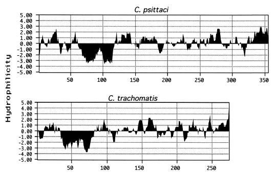
Comparison of IncA proteins from C. psittaci and C. trachomatis by hydropathy plot analysis. A hydropathy profile of each protein shows a unique bilobed hydrophobic domain in the N-terminal half. Profiles were determined by the algorithm developed by Kyte and Doolittle (9), with a window size of seven amino acids. The vertical axis displays relative hydrophilicity, with negative scores indicating relative hydrophobicity.
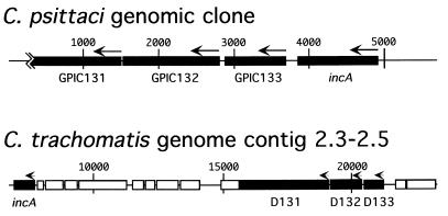
ORF map of the chromosomal region surrounding incA in C. psittaci and C. trachomatis. ORFs 131, 132, 133, and incA are labeled. Note the scale difference between the maps. ORF 133 is immediately downstream of incA in C. psittaci, whereas it is upstream and separated by at least 10 kb in C. trachomatis. Base pairs are indicated above each map, and arrows indicate the direction of transcription. The ORF designation is preserved from the C. trachomatis serovar D genome database designations. Pustell protein matrix analysis was used to confirm that GPIC131 and GPIC132 correspond to D131 and D132, respectively.
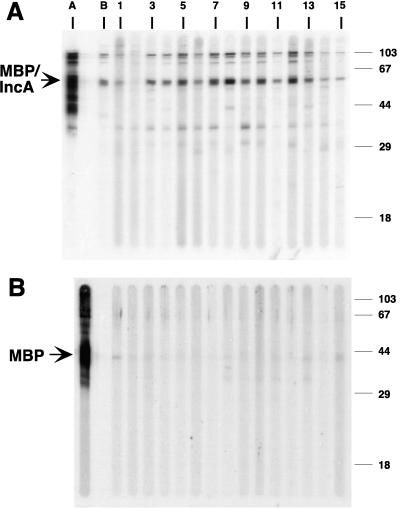
Preparative immunoblot analysis of a purified MBP-C. trachomatis IncA fusion protein (A) and purified MBP (B), each probed with antisera from chlamydia-infected patients and monkeys. Lane A, anti-MBP; lane B, monkey convalescent-phase sera; lanes 1 and 2, sera from C. pneumoniae-infected patients; lanes 3 to 13, sera from C. trachomatis-infected patients; lanes 14 and 15, negative control sera.
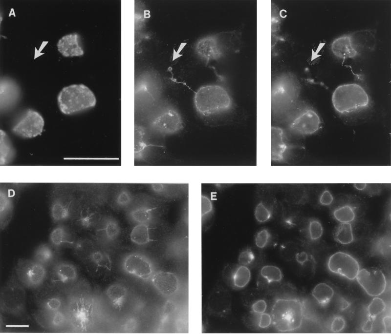
Immunofluorescence microscopy with anti-IncA demonstrating that IncA is localized to the inclusion membrane in C. trachomatis-infected cells. Serovar L2-infected HeLa cells were fixed in methanol 25 h postinfection and stained with anti-major outer membrane protein (A) and/or anti-MBP-IncA (B to E). Panels A to C represent a single image, with panel C photographed in a different focal plane. Note the fibers extending between the two inclusions in different cells as well as from one infected cell to an apparently uninfected cell (uninfected cell at tip of arrow). Note also the antigenic fibers extending from several inclusions in one focal plane (D) and IncA in inclusions at different stages of maturation in another focal plane (E). Bars in panels A and D represent 10 μm for panels A to C and panels D and E, respectively.
References
-
- Bannantine J P, Rockey D D, Hackstadt T. Tandem genes of Chlamydia psittaci that encode proteins localized to the inclusion membrane. Mol Microbiol. 1998;28:1017–1026. - PubMed
Publication types
MeSH terms
Substances
Associated data
- Actions
Grants and funding
LinkOut - more resources
Full Text Sources
Other Literature Sources
Molecular Biology Databases
