. 1998 Nov 24;95(24):14028-33.
doi: 10.1073/pnas.95.24.14028.

Transgenic cattle produced by reverse-transcribed gene transfer in oocytes

Affiliations

Transgenic cattle produced by reverse-transcribed gene transfer in oocytes

A W Chan et al. Proc Natl Acad Sci U S A. .

Abstract

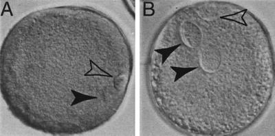
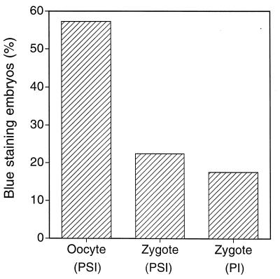
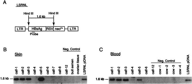
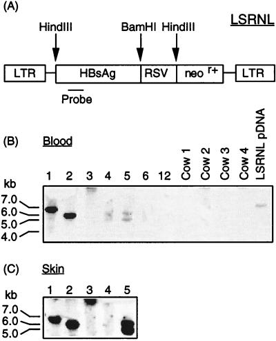
A critical requirement for integration of retroviruses, other than HIV and possibly related lentiviruses, is the breakdown of the nuclear envelope during mitosis. Nuclear envelope breakdown occurs during mitotic M-phase, the envelope reforming immediately after cell division, thereby permitting the translocation of the retroviral preintegration complex into the nucleus and enabling integration to proceed. In the oocyte, during metaphase II (MII) of the second meiosis, the nuclear envelope is also absent and the oocyte remains in MII arrest for a much longer period of time compared with M-phase in a somatic cell. Pseudotyped replication-defective retroviral vector was injected into the perivitelline space of bovine oocytes during MII. We show that reverse-transcribed gene transfer can take place in an oocyte in MII arrest of meiosis, leading to production of offspring, the majority of which are transgenic. We discuss the implications of this mechanism both as a means of production of transgenic livestock and as a model for naturally occurring recursive transgenesis.


Figures

Figure 1
Figure 1
Bovine oocytes matured in vitro demonstrate distinctive nuclear configuration compared with zygotes at the pronuclear stage. Oocytes were fixed 18 hr after placing in maturation medium and zygotes were fixed 18 hr postsemen addition, in three parts ethanol/one part acetic acid for 24 hr, before acid-orcein staining (1% orcein stain in 40% acetic acid in H2O). (A) After induction of maturation, the oocyte undergoes germinal vesicle (4N) breakdown, the chromatin condenses, the first meiotic division occurs, and the first polar body, containing half of the genome (2N), is extruded. The remaining condensed, diploid chromatin is aligned at the second metaphase plate (arrowhead). Chromatin is not enclosed in a nuclear membrane and is located next to the first polar body. The polar body also has intense chromatin, which is enclosed in a plasma membrane during autosome segregation (open arrowhead). The oocyte remains arrested in MII until the completion of meiosis, which is heralded by the extrusion of the second polar body (1N) induced by fertilization. (B) After fertilization, the oocyte progresses from MII to interphase. Zygote contains both maternal and paternal pronuclei enclosed by a nuclear envelope (arrowhead). The second polar body is located next to the maternal pronucleus with distinctive chromatin staining (open arrowhead). In addition to the difference in nuclear configuration, oocytes have very condensed chromatin and intense chromatin staining, whereas zygotes have dispersed chromatin with less intense staining.
Figure 2
Figure 2
Gene transfer efficiency of LRgeoL-(VSV-G) in bovine oocytes and zygotes. Gene transfer efficiency was evaluated by X-gal staining of embryos 4 days postsemen addition. Pronuclear injection of linearized SV-lac into zygotes was used as a control. Infection with LRgeoL-(VSV-G) was achieved by PSI at the indicated time points. (A) Oocytes at 20 hr after placing in maturation medium were infected by LRgeoL-(VSV-G) PSI. Four hours after infection, oocytes were incubated with thawed semen. X-gal staining was observed in 56% (178/316) of infected embryos. Zygotes at 18 hr postsemen addition were infected by LRgeoL-(VSV-G) PSI. X-gal staining was observed in 22% (49/226) of infected embryos. Zygotes at 18 hr postsemen addition were pronuclear injected with SV-lac, and 17% (25/144) of microinjected embryos were stained with X-gal. Cytoplasmic expression, in the case of pronuclear microinjection, does not differentiate between integration and extrachromosomal expression.
Figure 3
Figure 3
PCR analysis to detect neor+ (A) and HBsAg (B) transgenes in DNA extracts of whole blood (mesoderm) and skin (ectoderm) of calves derived from embryos infected with VSV-G-pseudotyped vector. (A and B) Lanes 1–5 are DNA from blood samples and lanes 6–10 are DNA from skin tissue of transgenic calves (lane 1: zygote treatment group, male; lane 2: oocyte treatment group, female; lane 3: oocyte treatment group, female; lane 4: oocyte reatment group, female; lane 5; oocyte treatment group, male.). Lanes 4 and 5 were from twins resulting from implantation of two embryos and the birth of phenotypically distinct calves of different breeds. The negative controls in lanes 11–13 comprise DNA from a blood sample from calf 12, which was a naturally conceived nontransgenic calf, commercial bull semen and ovarian tissue from a cow. Lane 14 contains the plasmid DNA of LSRNL, and lane 15 is a water control. Tissue samples from blood and skin show positive signal with both primer sets, but none of the negative controls show the presence of transgene. Differences in intensity of signal do not reflect copy numbers of the transgene inserted.
Figure 4
Figure 4
Southern blot analysis of HindIII-digested genomic DNA (A) to detect the 1.6-kb fragment from the HBsAg gene in blood (B) and skin (C) tissue of calves. (B) Lanes 1–5, transgenic calves. Lane 6, nontransgenic calf born after VSV-G-pseudotyped vector injection. Lanes 7 and 8, nontransgenic aborted fetuses. Negative controls include calf 12, which was a naturally conceived nontransgenic calf, ovarian tissue from a cull cow and commercial bull semen. (C) Lanes 1–5, transgenic calves. Lane 6, nontransgenic calf. Negative controls, calf 12 (naturally conceived nontransgenic). Cows 1–4 were the recipients carrying the transgenic calves. HindIII-digested pLSRNL DNA was included as a positive control.
Figure 5
Figure 5
Detection of chromosomal integration of HBsAg gene by Southern blot hybridization of the BamHI-digested genomic DNA from blood and skin tissue of calves. (A) Digestion sites of pLSRNL. Various-sized fragments were produced from different calves that were different from the linearized plasmid DNA control, demonstrating the successful unique insertion of the transgene into the host cell genome of each calf. (B) Blood—lanes 1–5, transgenic calves. Lane 6, nontransgenic calves born after VSV-G-pseudotyped vector injection. The negative controls in lanes 7–11 comprise calf 12, a naturally conceived nontransgenic calf, and blood samples from recipient cows (nos. 1–4) that carried the transgenic embryos. Lane 12 is the plasmid DNA of LSRNL digested by BamHI. (C) Skin—lanes 1–5, transgenic calves that show the same integration pattern as the corresponding blood samples.

References

    1. Wall R J. Theriogenology. 1996;45:57–68.
    1. Jaenisch R. Proc Natl Acad Sci USA. 1976;73:1260–1264. - PMC - PubMed
    1. Jaenisch R. Cell. 1980;19:181–188. - PubMed
    1. Haskell R E, Bowen R A. Mol Reprod Dev. 1995;40:386–390. - PubMed
    1. Kim T, Leibfried-Rutledge M L, First N L. Anim Biotechnol. 1993;4:53–69.

Publication types

MeSH terms

Substances

LinkOut - more resources