

doi: 10.1073/pnas.95.24.14066.
DNA end-joining catalyzed by human cell-free extracts
Affiliations
- PMID: 9826654
- PMCID: PMC24327
- DOI: 10.1073/pnas.95.24.14066
Item in Clipboard
DNA end-joining catalyzed by human cell-free extracts
Proc Natl Acad Sci U S A.
.
Abstract
Mammalian cells defective in DNA end-joining are highly sensitive to ionizing radiation and are immunodeficient because of a failure to complete V(D)J recombination. By using cell-free extracts prepared from human lymphoblastoid cell lines, an in vitro system for end-joining has been developed. Intermolecular ligation was found to be accurate and to depend on DNA ligase IV/Xrcc4 and requires Ku70, Ku86, and DNA-PKcs, the three subunits of the DNA-activated protein kinase DNA-PK. Because these activities are involved in the cellular resistance to x-irradiation and V(D)J recombination, the development of this in vitro system provides an important advance in the study of the mechanism of DNA end-joining in human cells.
Figures
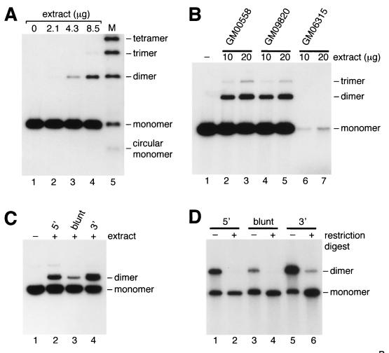
DNA end-joining catalyzed by human cell-free extracts. (A) Protein extracts from GM00558 were incubated with 5′-32P-end-labeled BsaI-linearized pDEA-7Z DNA as described in Materials and Methods (lanes 1–4). Lane 5, DNA ligation ladder. (B) Extracts of three lymphoblastoid cell lines were analyzed for their ability to promote end-joining under standard assay conditions. (C) End-joining catalyzed by GM00558 extract (68 μg) was analyzed by using uniformly 32P-labeled pFB585 DNA (100 ng) linearized with BsaI (lanes 1 and 2), EcoRV (lane 3), or KpnI (lane 4). (D) Ligation products from reactions similar to those shown in C were purified by gel electrophoresis and treated with (+) or without (−) BsaI (lanes 1 and 2), EcoRV (lanes 3 and 4), or KpnI (lanes 5 and 6).
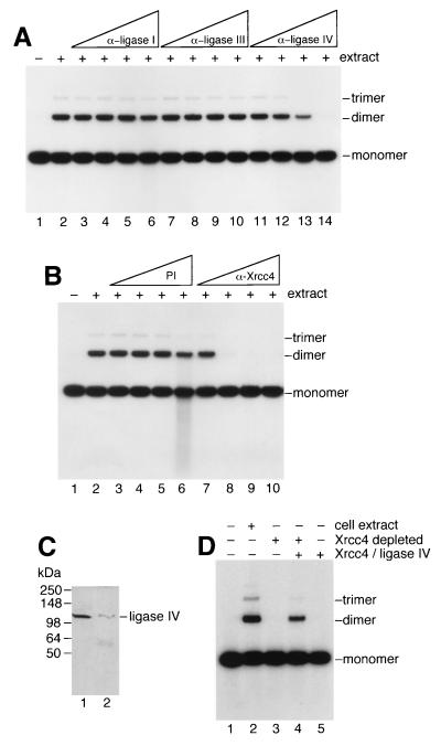
Involvement of DNA ligase IV and Xrcc4 in end-joining. (A) End-joining reactions were carried out as described for Fig. 1 by using GM00558 extract (34 μg) but contained polyclonal antisera (1 μl neat, lanes 6, 10, 14; diluted 1/5, lanes 5, 9, 13; diluted 1/25, lanes 4, 8, 12; or diluted 1/125, lanes 3, 7, 11) against DNA ligases I, III or IV as indicated. Lanes: 1, no extract; 2, complete reaction but without antiserum. Extract was incubated for 30 min on ice with antiserum, then was transferred to 37°C for 10 min followed by the addition of 32P-end-labeled DNA (10 ng). After 1-hour incubation, DNA products were analyzed by agarose gel electrophoresis. (B) Reactions were carried out as described in A by using preimmune serum (lanes 3–6) or anti-Xrcc4 antiserum (lanes 7–10). (C) Immunodepletion of Xrcc4/Ligase IV determined by Western analysis. Lanes 1 and 2, extract before and after immunodepletion of Xrcc4/DNA ligase IV by using anti-Xrcc4 antiserum. (D) Analysis of end-joining before and after Xrcc4/ligase IV immunodepletion. The presence of cell extract or immunodepleted extract is indicated. Purified Xrcc4/ligase IV was added where shown. End-joining assays were carried out as described for Fig. 1.
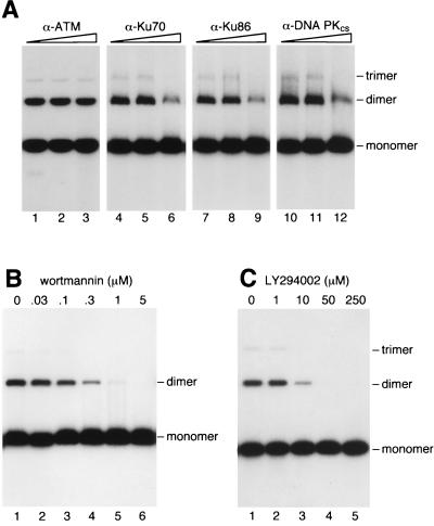
Involvement of DNA-PK in end-joining. (A) End-joining reactions contained GM00558 cell-free extract (20 μg) and 32P-labeled linear DNA. Reactions were supplemented with dilutions of antisera and analyzed as described for Fig. 2. (B and C) Inhibition of end-joining by the DNA-PK inhibitors wortmannin and LY294002.
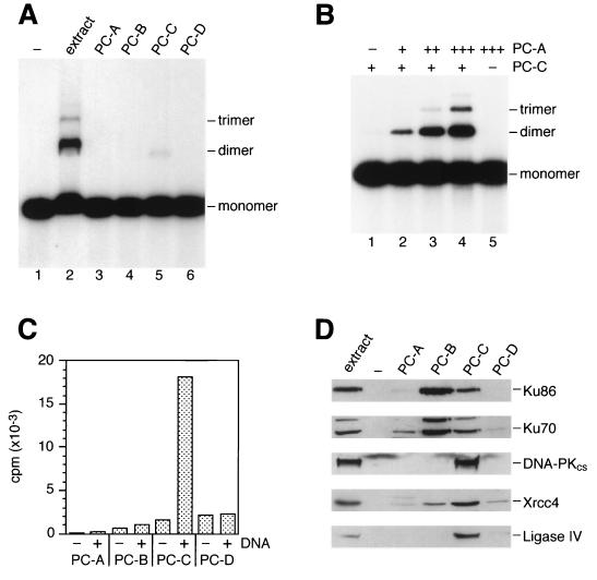
Fractionation of end-joining activity. (A) GM00558 extract was fractionated by phosphocellulose chromatography as described in Materials and Methods. End-joining assays then were carried out by using crude extract (20 μg) or fractions PC-A, PC-B, PC-C, and PC-D (2 μl of each) as indicated. (B) Fraction PC-C (2.2 μg) was mixed on ice with 0, 0.3, 1.0, or 3.0 μg of fraction PC-A (lanes 1–4). After 5 min at 37°C, 32P-labeled linear DNA was added, and end-joining reactions were carried out for 1 hour. Lane 5, reaction contained PC-A (3 μg) only. (C) DNA-PK activity was measured by incubating 3.3 μg of the four fractions with biotinylated peptide substrate in the absence (−) and presence (+) of sonicated calf thymus DNA by using the Promega SignaTECT DNA-PK assay system. (D) Western blotting. Membranes probed with antisera against Ku70, Ku86, and DNA-PKcs contained 8 μg of total protein per gel lane, and those probed with anti-Xrcc4 and anti-ligase IV contained 30 μg of protein per lane.
References
-
- Thompson L H, Jeggo P A. Mutat Res. 1995;337:131–137. - PubMed
-
- Li Z Y, Otevrel T, Gao Y J, Cheng H L, Seed B, Stamato T D, Taccioli G E, Alt F W. Cell. 1995;83:1079–1089. - PubMed
-
- Critchlow S E, Bowater R P, Jackson S P. Curr Biol. 1997;7:588–598. - PubMed
-
- Grawunder U, Wilm M, Wu X T, Kulesza P, Wilson T E, Mann M, Lieber M R. Nature (London) 1997;388:492–495. - PubMed
Publication types
MeSH terms
Substances
LinkOut - more resources
Full Text Sources
Other Literature Sources
Molecular Biology Databases
Research Materials
Miscellaneous
