

Ribosome display efficiently selects and evolves high-affinity antibodies in vitro from immune libraries
- PMID: 9826665
- PMCID: PMC24338
- DOI: 10.1073/pnas.95.24.14130
Ribosome display efficiently selects and evolves high-affinity antibodies in vitro from immune libraries
Abstract
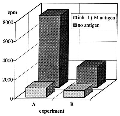
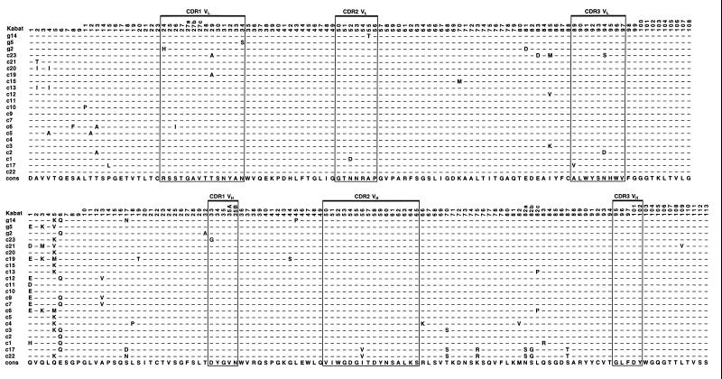
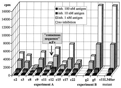
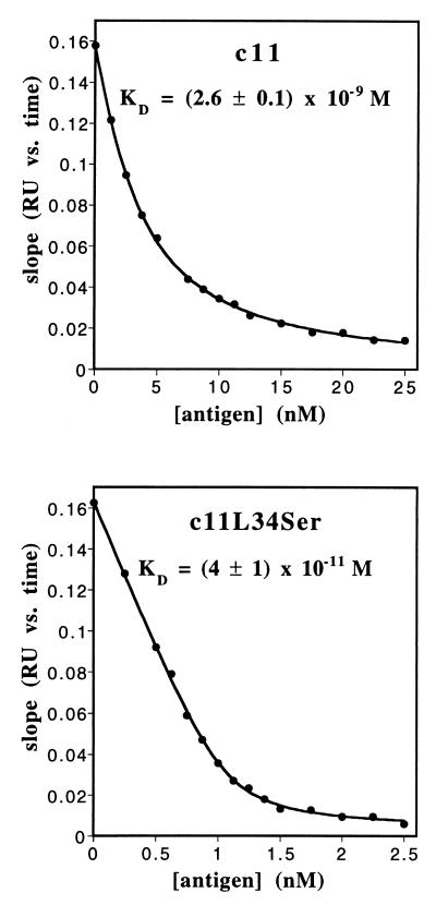
Ribosome display was applied for affinity selection of antibody single-chain fragments (scFv) from a diverse library generated from mice immunized with a variant peptide of the transcription factor GCN4 dimerization domain. After three rounds of ribosome display, positive scFvs were isolated and characterized. Several different scFvs were selected, but those in the largest group were closely related to each other and differed in 0 to 5 amino acid residues with respect to their consensus sequence, the likely common progenitor. The best scFv had a dissociation constant of (4 +/- 1) x 10(-11) M, measured in solution. One amino acid residue in complementarity determining region L1 was found to be responsible for a 65-fold higher affinity than the likely progenitor. It appears that this high-affinity scFv was selected from the mutations occurring during ribosome display in vitro, and that this constitutes an affinity maturation inherent in this method. The in vitro-selected scFvs could be functionally expressed in the Escherichia coli periplasm with good yields or prepared by in vitro refolding. Thus, ribosome display can be a powerful methodology for in vitro library screening and simultaneous sequence evolution.
Figures
Comment in
-
mRNA display: diversity matters during in vitro selection.Proc Natl Acad Sci U S A. 2001 Apr 24;98(9):4825-6. doi: 10.1073/pnas.091101698. Proc Natl Acad Sci U S A. 2001. PMID: 11320229 Free PMC article. No abstract available.
References
-
- Saffhill R, Schneider-Bernloehr H, Orgel L E, Spiegelman S. J Mol Biol. 1970;51:531–539. - PubMed
-
- Gold L, Polisky B, Uhlenbeck O, Yarus M. Annu Rev Biochem. 1995;64:763–797. - PubMed
-
- Irvine D, Tuerk C, Gold L. J Mol Biol. 1991;222:739–761. - PubMed
-
- Dower W J, Cwirla S E. In: Guide to Electroporation and Electrofusion. Chang D C, Chassy B M, Saunders J A, Sowers A E, editors. San Diego: Academic; 1992. pp. 291–301.
Publication types
MeSH terms
Substances
LinkOut - more resources
Full Text Sources
Other Literature Sources
