

Insulin depletion leads to adipose-specific cell death in obese but not lean mice
- PMID: 9826672
- PMCID: PMC24345
- DOI: 10.1073/pnas.95.24.14168
Insulin depletion leads to adipose-specific cell death in obese but not lean mice
Abstract
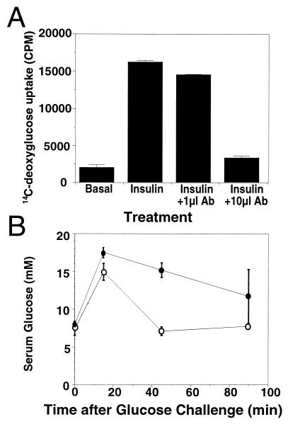
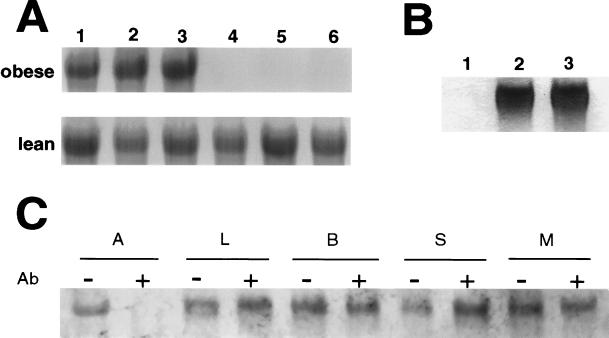
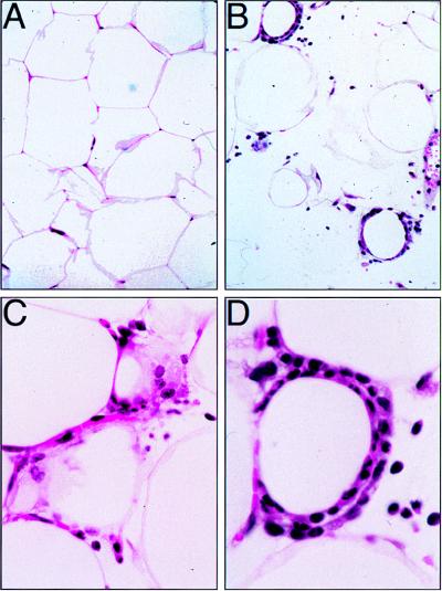
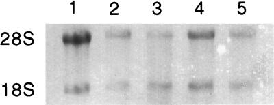
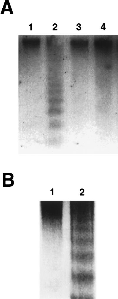
Mutation of the obese gene produces obesity, hyperinsulinemia, and compensatory "overexpression" of the defective gene. As insulin activates obese gene expression, it seemed possible that hyperinsulinemia might be responsible for overexpression of the gene. To address this question we rapidly neutralized circulating insulin by injection of an insulin antibody. Unexpectedly, insulin depletion in obese (ob/ob or db/db) mice caused massive adipose RNA degradation confirmed by histological analysis to result from adipocyte cell death by a largely necrotic mechanism. This effect was not observed in lean littermates and was completely corrected by coadministration of insulin. Comparison of multiple tissues demonstrated that the effect was restricted to adipose tissue. Insulin depletion in obese mice by administration of streptozotocin also led to cell death, but this death was less extensive and appeared to be apoptotic in mechanism. Thus insulin may promote the survival side of the physiological balance between adipocyte survival and death.
Figures
Similar articles
-
Regulated expression of the obese gene product (leptin) in white adipose tissue and 3T3-L1 adipocytes.Proc Natl Acad Sci U S A. 1995 Sep 26;92(20):9034-7. doi: 10.1073/pnas.92.20.9034. Proc Natl Acad Sci U S A. 1995. PMID: 7568067 Free PMC article.
-
Angiotensinogen gene expression in adipose tissue: analysis of obese models and hormonal and nutritional control.Am J Physiol. 1997 Jul;273(1 Pt 2):R236-42. doi: 10.1152/ajpregu.1997.273.1.R236. Am J Physiol. 1997. PMID: 9249555
-
Increased sensitivity of the genetically obese mouse to corticosterone.Am J Physiol. 1987 Feb;252(2 Pt 1):E202-8. doi: 10.1152/ajpendo.1987.252.2.E202. Am J Physiol. 1987. PMID: 3548420
-
Multihormonal control of ob gene expression and leptin secretion from cultured human visceral adipose tissue: increased responsiveness to glucocorticoids in obesity.J Clin Endocrinol Metab. 1998 Mar;83(3):902-10. doi: 10.1210/jcem.83.3.4644. J Clin Endocrinol Metab. 1998. PMID: 9506746 Review.
-
Interrelationships between obesity and diabetes.Proc Nutr Soc. 1991 Dec;50(3):605-18. doi: 10.1079/pns19910074. Proc Nutr Soc. 1991. PMID: 1809969 Review. No abstract available.
Cited by
-
Differential roles of CIDEA and CIDEC in insulin-induced anti-apoptosis and lipid droplet formation in human adipocytes.J Lipid Res. 2010 Jul;51(7):1676-84. doi: 10.1194/jlr.M002147. Epub 2010 Feb 14. J Lipid Res. 2010. PMID: 20154362 Free PMC article.
-
Differential regulation of CIDEA and CIDEC expression by insulin via Akt1/2- and JNK2-dependent pathways in human adipocytes.J Lipid Res. 2011 Aug;52(8):1450-60. doi: 10.1194/jlr.M012427. Epub 2011 Jun 2. J Lipid Res. 2011. PMID: 21636835 Free PMC article.
-
A novel JNK2/SREBP-1c pathway involved in insulin-induced fatty acid synthesis in human adipocytes.J Lipid Res. 2013 Jun;54(6):1531-1540. doi: 10.1194/jlr.M031591. Epub 2013 Mar 19. J Lipid Res. 2013. PMID: 23515281 Free PMC article. Clinical Trial.
-
Lack of Wdr13 gene in mice leads to enhanced pancreatic beta cell proliferation, hyperinsulinemia and mild obesity.PLoS One. 2012;7(6):e38685. doi: 10.1371/journal.pone.0038685. Epub 2012 Jun 8. PLoS One. 2012. PMID: 22715406 Free PMC article.
-
Hypotension, lipodystrophy, and insulin resistance in generalized PPARgamma-deficient mice rescued from embryonic lethality.J Clin Invest. 2007 Mar;117(3):812-22. doi: 10.1172/JCI28859. Epub 2007 Feb 15. J Clin Invest. 2007. PMID: 17304352 Free PMC article.
References
Publication types
MeSH terms
Substances
Grants and funding
LinkOut - more resources
Full Text Sources
Other Literature Sources
Medical
Molecular Biology Databases
Research Materials
Miscellaneous
