

doi: 10.1073/pnas.95.24.14179.
An essential role of phosphatidylinositol 3-kinase in myogenic differentiation
Affiliations
- PMID: 9826674
- PMCID: PMC24347
- DOI: 10.1073/pnas.95.24.14179
Item in Clipboard
An essential role of phosphatidylinositol 3-kinase in myogenic differentiation
Proc Natl Acad Sci U S A.
.
Abstract
The oncogene p3k, coding for a constitutively active form of phosphatidylinositol 3-kinase (PI 3-kinase; EC 2.7.1.137), strongly enhances myogenic differentiation in cultures of chicken-embryo myoblasts. It increases the size of the myotubes and induces elevated levels of the muscle-specific proteins MyoD, myosin heavy chain, creatine kinase, and desmin. Inhibition of PI 3-kinase activity with LY294002 or with dominant-negative mutants of PI 3-kinase interferes with myogenic differentiation and with the induction of muscle-specific genes. PI 3-kinase is therefore an upstream mediator for the expression of the muscle-specific genes and is both necessary and rate-limiting for the process of myogenesis.
Figures
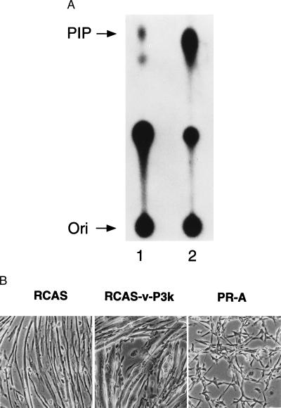
(A) Increase of PI 3-kinase activity in CEM expressing v-P3k. CEM were prepared as described (27) and were infected with the retroviral vector RCAS (lane 1) or with RCAS-v-P3k (lane 2). The cells were cultured for 2 days in MG medium, followed by three days in MD medium as described in Materials and Methods. PI 3-kinase activity was assayed in vitro as described (29) PIP, phosphatidylinositol 3-phosphate; Ori, origin of the chromatogram. (B) Enhancement of myogenic differentiation by v-P3k. CEM were infected with the RCAS vector, RCAS-v-P3k, or Prague strain Rous sarcoma virus subgroup A (PR-A) and cultured as above. Representative fields were photographed 5 days postinfection (×6.5 objective, phase-contrast).
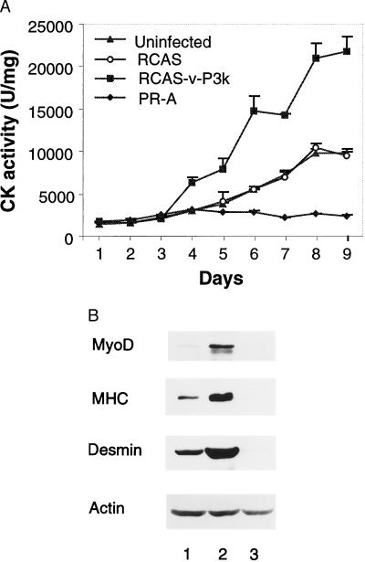
(A) Induction of creatine kinase (CK) activity by v-P3k. Noninfected CEM or CEM infected with RCAS viruses or PR-A as described in Fig. 1 were kept in MG medium for 2 days and thereafter were maintained in MD medium. Cells were harvested at daily intervals, and CK activity was assayed by using a commercial kit (Sigma). Mean ± SE values were obtained from two (uninfected and PR-A-infected) or three (RCAS and RCAS-v-P3k-infected) independent experiments (two replicate plates per experiment). The CK activity is expressed in μmol of creatine formed per min per mg of total protein extract (U/mg). (B) Induction of muscle-specific proteins by v-P3k. Total proteins were prepared from CEM at day 8 postinfection with RCAS vector (lane 1), RCAS-v-P3k (lane 2), or PR-A (lane 3). Muscle-specific protein levels were analyzed by immunoblot assay by using antibodies specific for MyoD (Santa Cruz Biotechnology), myosin heavy chain (MHC), and desmin (ICN). Actin served as ubiquitously expressed control was detected with an antibody from Sigma. Similar results were obtained in three independent experiments.
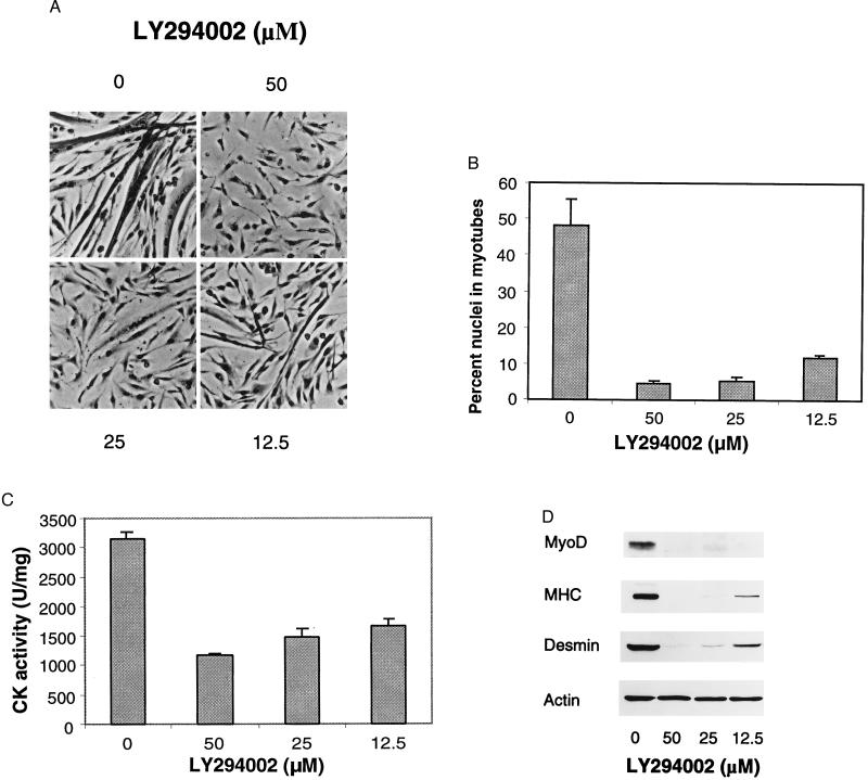
(A) Inhibition of myoblast differentiation by the PI 3-kinase-specific inhibitor LY294002. CEM were cultured in MG medium for 1 day after seeding, and continued to grow for 3 days in MD medium containing the indicated concentrations of LY294002 or dimethyl sulfoxide solvent. Cells were stained on day 4 with the LeukoStat kit (Fisher) to distinguish the cytoplasm and the nucleus, and representative fields were photographed (×16 objective, brightfield illumination). (B) Inhibition of multinucleated myotube formation by LY294002. Cells on day 4 were stained as above, and counted under ×16 objective lens to determine the percentage of nuclei in myotubes (containing at least three nuclei per cell). Three independent plates with three random fields per plate were used. (C) Inhibition of CK activity by LY294002. CK activity on day 4 was determined in two experiments with three replicate plates in the presence of LY294002 as indicated. (D) Inhibition of muscle-specific gene expression by LY294002. Total protein was extracted from CEM treated for 3 days with LY294002 as indicated and processed for immunoblot assay as described in Fig. 2B.
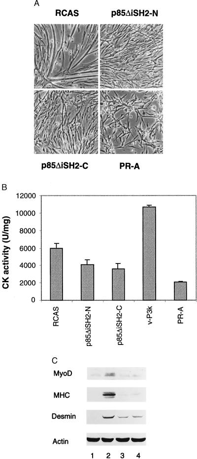
(A) Dominant-negative PI 3-kinase constructs inhibit myogenesis. CEM were infected with RCAS, PR-A, or RCAS viruses expressing two dominant-negative mutants of the regulatory subunit of PI 3-kinase, p85ΔiSH2-N and p85ΔiSH2-C (43). The cells were cultured in MG medium for 3 days postinfection, followed by 2 days in MD medium to induce myoblast differentiation. Representative fields were photographed on day 5 (×6.5 objective, phase-contrast). (B) Dominant-negative PI 3-kinase mutants interfere with the induction of CK. CK activity was determined on day 6 in CEM cultures infected as described above or infected with RCAS-v-P3k as a positive control. Mean ± SE values were from two experiments with three replicate assays. (C) Dominant-negative PI 3-kinase mutants inhibit the expression of muscle-specific proteins. Muscle-specific protein levels were studied in the cells on day 6 postinfection with PR-A (lane 1), RCAS (lane 2), RCAS-p85ΔiSH2-N (lane 3), or RCAS-p85ΔiSH2-C (lane 4) viruses and assayed by immunoblot as in Fig. 2B. Similar results were obtained in three replicate experiments.
References
-
- Davis R L, Weintraub H, Lassar A B. Cell. 1987;51:987–1000. - PubMed
-
- Weintraub H, Davis R, Tapscott S, Thayer M, Krause M, Benezra R, Blackwell T K, Turner D, Rupp R, Hollenberg S, et al. Science. 1991;251:761–766. - PubMed
-
- Lassar A, Munsterberg A. Curr Opin Cell Biol. 1994;6:432–442. - PubMed
-
- Lassar A B, Buskin J N, Lockshon D, Davis R L, Apone S, Hauschka S D, Weintraub H. Cell. 1989;58:823–831. - PubMed
-
- Molkentin J D, Olson E N. Curr Opin Genet Dev. 1996;6:445–453. - PubMed
Publication types
MeSH terms
Substances
Grants and funding
LinkOut - more resources
Full Text Sources
Research Materials
Miscellaneous
