. 1998 Nov 24;95(24):14208-13.
doi: 10.1073/pnas.95.24.14208.

Testatin: a cystatin-related gene expressed during early testis development

Affiliations

Testatin: a cystatin-related gene expressed during early testis development

V Töhönen et al. Proc Natl Acad Sci U S A. .

Abstract

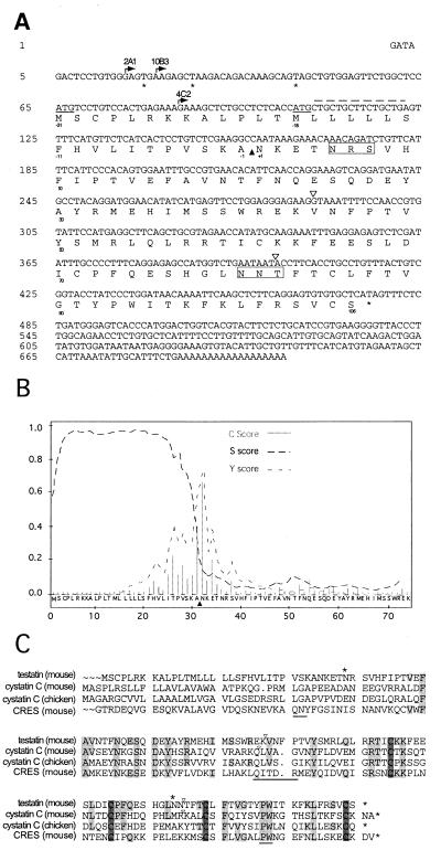
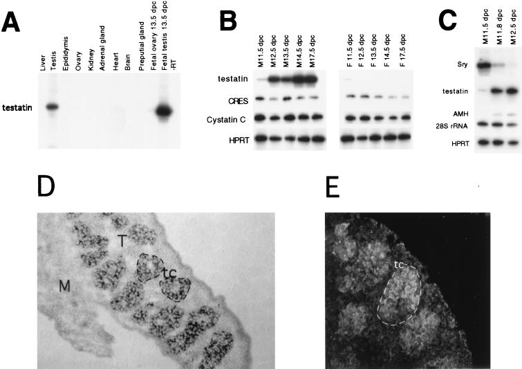
To isolate genes involved in morphogenic aspects of testis development, and which may act in cell signaling pathways downstream of the testis-determining gene Sry, we have developed a modified mRNA differential display method named signal peptide differential display. It was used to target those genes that encode proteins having a signal peptide sequence. By using this method, we isolated a gene named testatin. This gene was found to be related to a group of genes that encodes cysteine protease inhibitors known as cystatins. Cystatins and their target proteases have been associated with tumor formation and metastasis, but also are involved in natural tissue remodeling events such as bone resorption and embryo implantation. We show that testatin expression is restricted to fetal gonads and adult testis. Furthermore, testatin is expressed during testis cord formation in pre-Sertoli cells, believed to be the site of Sry action, at a time immediately after the peak of Sry expression. This finding suggests that testatin might be activated by transcription factors that are known to orchestrate the early testis development pathway. This gene therefore represents one of the putative downstream targets likely to have an essential role in tissue reorganization during early testis development.


Figures

Figure 1
Figure 1
(A) Schematic diagram comparing the SPDD and the classic DD. (B) An SPDD screen using total RNA from 13.5 dpc male and female fetal mouse gonads+mesonephros identified a male-specific fragment (M12). RTs were performed by using four different decamer primers 5*13, 5*14, 5*15, and 5*16. By PCR, cDNAs were amplified by using the different decamers in combination with the signal peptide primer in the presence of [α-33P] dATP for 28 cycles. cDNA bands were resolved on a denaturing polyacrylamide gel and exposed to x-ray film. M12 was isolated, cloned, and sequenced.
Figure 2
Figure 2
(A) Nucleotide and predicted amino acid sequence of the testatin gene. The start sites for the three testatin cDNA clones—2A1 (nucleotide 17), 10B3 (nucleotide 22), and 4C2 (nucleotide 85)—are indicated by arrows. The two predicted translation initiation methionine codons, ATG, are underlined (nucleotides 65–57 and 104–106), and stop codons are indicated by ∗. Hatched line shows the potential binding site for the signal peptide primer. The predicted signal peptide cleavage site is indicated by a filled arrow. The boundaries between exons and introns are indicated by open triangles. Potential N-glycosylation sites are marked with open boxes. (B) Prediction of the testatin signal peptide cleavage site. The amino acid sequence deduced from testatin was analyzed to determine the cleavage site between the signal sequence and the mature protein by using the SignalP V1.1 World Wide Web Server (14). The black arrow indicates the most probable cleavage site. (C) Alignment of the derived testatin amino acid sequence with mouse cystatin C, chicken cystatin C, and CRES. Putative signal peptides are indicated in pale gray boxes. The numbering follows the predicted testatin sequence. Gray boxes show homologous regions between testatin and the cystatins. Dark gray boxes are the aligned cysteine residues. The marked amino acids (∗) indicate possible N-glycosylation sites of the testatin protein. The lower black line lies over the three domains thought to be critical to the cystatins for cysteine protease inhibitory activity. The third domain, PW, is common for testatin, cystatins, and CRES.
Figure 3
Figure 3
Testatin is expressed specifically in the male gonad soon after Sry is expressed and later in adult testis. (A) Semiquantitative RT-PCR analysis of testatin mRNA expression in adult mouse organs. (B) Time-course analysis of testatin, cystatin C, CRES, and HPRT expression in developing gonads, from 11.5 to 17 dpc, using semiquantitative RT-PCR. (C) Time-course analysis of testatin, Sry, AMH, and HPRT expression during the initial phase of testis development, 11.5 to 12.5 dpc, using semiquantitative RT-PCR. (D) In situ analysis using a digoxigenin-labeled antisense testatin probe on sections from fetal testis (T)+mesonephros (M), 13.5 dpc. Testatin expression is located to the Sertoli cells in the testis cords (tc). (E) Immunostaining of testatin protein in fetal testis, 13.5 dpc, using a testatin-specific antibody.

Similar articles

Cited by

References

    1. Nordqvist K, Lovell-Badge R. Nat Genet. 1994;7:7–9. - PubMed
    1. Gubbay J, Collignon J, Koopman P, Capel B, Economou A, Münsterberg A, Vivian N, Goodfellow P, Lovell-Badge R. Nature (London) 1990;346:245–250. - PubMed
    1. Sinclair A H, Berta P, Palmer M S, Hawkins J R, Griffiths B L, Smith M J, Foster J W, Frischauf A M, Lovell-Badge R, Goodfellow P N. Nature (London) 1990;346:240–244. - PubMed
    1. Koopman P, Gubbay J, Vivian N, Goodfellow P, Lovell-Badge R. Nature (London) 1991;351:117–121. - PubMed
    1. Capel B. Annu Rev Physiol. 1998;60:497–523. - PubMed

Publication types

MeSH terms

Associated data