. 1998 Nov 24;95(24):14250-5.
doi: 10.1073/pnas.95.24.14250.

A heterodimeric DNA polymerase: evidence that members of Euryarchaeota possess a distinct DNA polymerase

Affiliations

A heterodimeric DNA polymerase: evidence that members of Euryarchaeota possess a distinct DNA polymerase

I K Cann et al. Proc Natl Acad Sci U S A. .

Abstract

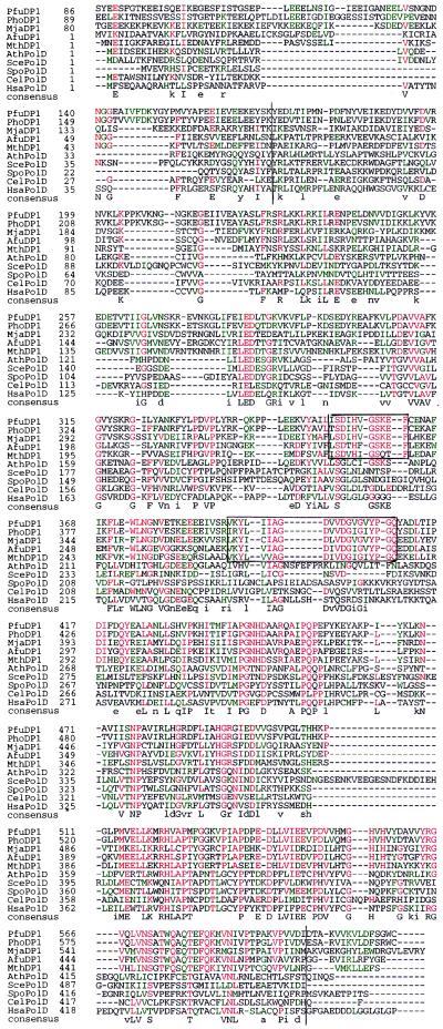
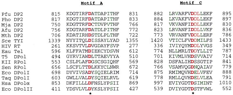
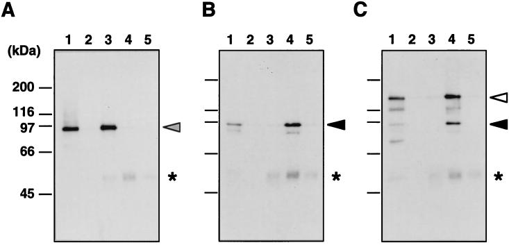
We describe here a DNA polymerase family highly conserved in Euryarchaeota, a subdomain of Archaea. The DNA polymerase is composed of two proteins, DP1 and DP2. Sequence analysis showed that considerable similarity exists between DP1 and the second subunit of eukaryotic DNA polymerase delta, a protein essential for the propagation of Eukarya, and that DP2 has conserved motifs found in proteins with nucleotide-polymerizing activity. These results, together with our previous biochemical analyses of one of the members, DNA polymerase II (DP1 + DP2) from Pyrococcus furiosus, implicate the DNA polymerases of this family in the DNA replication process of Euryarchaeota. The discovery of this DNA-polymerase family, aside from providing an opportunity to enhance our knowledge of the evolution of DNA polymerases, is a significant step toward the complete understanding of DNA replication across the three domains of life.


Figures

Figure 1
Figure 1
Amino acid sequence alignment of the euryarchaeal DP1 and eukaryotic Pol δ small subunit. The sequences were aligned with clustalw, version 1.7. The polymerases shown are from P. furiosus (Pfu), P. horikoshii (Pho), M. jannaschii (Mja), A. fulgidus (Afu), M. thermoautotrophicum (Mth), A. thaliana (Ath), S. cerevisiae (Sce), S. pombe (Spo), C. elegans (Cel), and H. sapiens (Hsa). The ORF names of the archaeal proteins obtained from their genome projects were Mja DP1 (MJ0702), Afu DP1 (AF1790), Mth DP1 (MTH1405), and Pho DP1 (PHBN023). Amino acid residues that are identical (red) or similar (green) in ≥50% of the positions are indicated. Upper case letters indicate consensus residues with identities at ≥50% positions, including, at least, one from each domain. Lowercase letters indicate the most frequent residues including, at least, one from each domain. The highly conserved regions from the eury archaeal group are boxed. Amino acids with similar properties are grouped as LIMV, AG, YWF, DEQN, KRH, and ST.
Figure 2
Figure 2
Amino acid sequence alignment showing two major conserved regions of polymerases, including the DP2 of Euryarchaeota. The sequences were aligned with clustalw, version 1.7. The asterisks indicate invariant residues. Amino acid residues that are identical (red) or similar (green) in ≥50% of the positions are indicated. The polymerases shown are from P. furiosus (Pfu), P. horikoshii (Pho), M. jannaschii (Mja), A. fulgidus (Afu), M. thermoautotrophicum (Mth), S. cerevisiae transposon TYI protein B (Sce TYI), HIV DNA polymerase polyprotein (HIV RT), E. aediculatus telomerase subunit (Eau Tel), bacteriophage T5 DNA polymerase (T5 DPol), bacteriophage KII RNA polymerase (KII Rpol), Sendai virus RNA polymerase β subunit (Sen Rpol), E. coli DNA polymerase I (Eco DPolI), T. aquaticus Pol I (Taq DPolI), H. sapiens DNA polymerase α (Hsa), and E. coli Pol II (Eco DpolII). The ORF names of the archaeal proteins obtained from their genome projects are Mja DP2 (MJ1630), Afu DP2 (AF1722), Mth DP2 (MTH1536), and Pho DP2 (PHBN021). Amino acids were classified as in Fig. 1.
Figure 3
Figure 3
Immunoprecipitation analysis of P. furiosus DNA polymerases. The total cell extracts were immunoprecipitated with three kinds of antiserum, anti-Pfu Pol I, anti-Pfu DP1, and anti-Pfu Pol II (DP1 + DP2). Immunoprecipitated fractions were separated by SDS/7.5% PAGE and then analyzed by Western blotting with anti-Pfu Pol I antiserum (A), anti-Pfu DP1 antiserum (B), and anti-Pfu Pol II antiserum (C). In each panel, lane 1 shows total cell extracts without immunoprecipitation; lane 2 shows total cell extracts precipitated with PBS; lane 3 shows total cell extracts precipitated with anti-Pfu Pol I; lane 4 shows total cell extracts precipitated with anti-Pfu DP1; and lane 5 shows total cell extracts precipitated with anti-PI-PfuI (negative control). The closed, open, and shaded arrowheads correspond to DP1, DP2, and Pol I, respectively. A nonspecific band (∗) found in all of the immunoprecipitated samples probably corresponds to IgG judging by its molecular size.

References

    1. Kornberg A, Baker T A. DNA Replication. 2nd Ed. New York: Freeman; 1992. pp. 169–182.
    1. Kelman Z, O’Donnell M. Annu Rev Biochem. 1995;64:171–200. - PubMed
    1. Stillman B. Cell. 1994;78:725–728. - PubMed
    1. Brush G S, Kelly T J. In: DNA Replication in Eukaryotic Cells. DePamphilis M L, editor. Plainview, NY: Cold Spring Harbor Lab. Press; 1996. pp. 1–43.
    1. Woese C R, Fox G E. Proc Natl Acad Sci USA. 1977;74:5088–5090. - PMC - PubMed

MeSH terms

Substances

LinkOut - more resources