. 1998 Nov 24;95(24):14314-9.
doi: 10.1073/pnas.95.24.14314.

Presentation of peptide antigens by mouse CD1 requires endosomal localization and protein antigen processing

Affiliations

Presentation of peptide antigens by mouse CD1 requires endosomal localization and protein antigen processing

S Tangri et al. Proc Natl Acad Sci U S A. .

Abstract

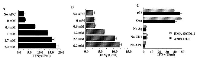
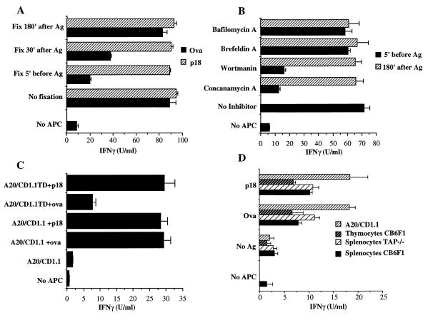
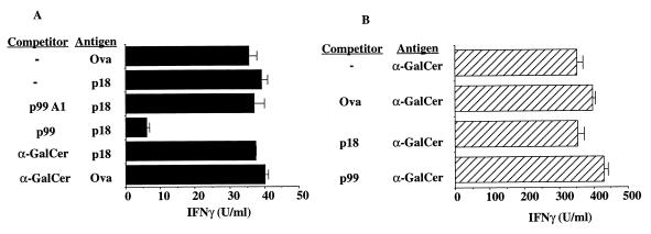
Mouse CD1(mCD1) molecules have been reported to present two types of antigens: peptides or proteins and the glycolipid alpha-galactosylceramide. Here, we demonstrate that a protein antigen, chicken ovalbumin (Ova), must be processed to generate peptides presented by mCD1 to CD8(+) T cells. The processing and mCD1-mediated presentation of chicken Ova depend on endosomal localization because inhibitors of endosomal acidification and endosomal recycling pathways block T cell reactivity. Furthermore, a cytoplasmic tail mutant of mCD1, which disrupts endosomal localization, has a greatly reduced capacity to present Ova to mCD1 restricted cells. Newly synthesized mCD1 molecules, however, are not required for Ova presentation, suggesting that molecules recycling from the cell surface are needed. Because of these data showing that mCD1 trafficks to endosomes, where it can bind peptides derived from exogenous proteins, we conclude that peptide antigen presentation by mCD1 is likely to be a naturally occurring phenomenon. In competition assays, alpha-galactosylceramide did not inhibit Ova presentation, and presentation of the glycolipid was not inhibited by excess Ova or the peptide epitope derived from it. This suggests that, although both lipid and peptide presentation may occur naturally, mCD1 may interact differently with these two types of antigens.


Figures

Figure 1
Figure 1
Cytokine release by mCD1-restricted T cells in response to Ova or Ova peptide. The response of T cells to APC pulsed with various concentrations of Ova (A) or p18 peptide (B) are shown. Reactive T cells were obtained from groups of four C57BL/6 mice immunized with 50–70 μg of plasmids encoding B7.1, Ova, and CD1.1. Spleen cells were harvested, were stimulated in vitro, and were restimulated with RMA-S/CD1.1 cells pulsed with indicated concentrations of antigen. The levels of IFN-γ were determined by ELISA. The IFN-γ produced by untransfected RMA-S APC pulsed with Ova or p18 was <2.5 units/ml. (C) The response of T cells from TAP−/− mice, generated and stimulated as described above, to Ova or p18 peptide presented by either A20 or RMA-S transfectants is shown. Representative data from one of three TAP−/− responder mice are shown.
Figure 2
Figure 2
Antigen processing and endosomal localization are required for Ova presentation by mCD1. (A) Fixed cells can present p18 peptide but not Ova. RMA-S/CD1.1 transfectants were fixed with 0.03% glutaraldehyde either before, or at different time points after, addition of antigens. The antigens were either 50 μg/ml Ova or 4 μg/ml p18 peptide. (B) RMA-S/CD1.1 transfectants were incubated with the indicated inhibitors: 20 nM Concanamycin A, 50 nM Bafilomycin, 200 nM Wortmannin, or 10 μg/ml Brefeldin A. Inhibitors were added either 5 min before, or 180 min after, addition of 50 μg/ml Ova. After 4 hr in the presence of the inhibitors, the APC were fixed and tested for their ability to stimulate Ova reactive T cells. (C) A20/CD1.1 and A20/CD1.1TD transfectants were pulsed either with 50 μg/ml Ova or 4 μg/ml p18 peptide for 3 hr, were irradiated, and were added to T cell stimulation assays. ELISAs were used to measure the IFN-γ levels after 3 days. In the case of A and B, the in vitro activated CTLs were derived from C57BL/6 mice whereas in C the CTLs were derived from CB6F1 mice. The control production of cytokine from untransfected cells pulsed with Ova, or T cells alone, in each case was <6 units/ml. These data are representative of one of six experiments. (D). Normal APC can present Ova to mCD1-restricted T cells. Ova-reactive T cells were generated from CB6F1 DNA immunized mice and were restimulated with Ova plus mCD1 RMA-S transfectants as described above. The representative data from one of six animals analyzed in this way is shown.
Figure 3
Figure 3
Lack of mutual competition between peptide and lipid antigens. (A) A lipid antigen does not compete for peptide recognition. A20/CD1.1 transfectants were pulsed with either 5.5 μM p99, 5.5 μM p99A1, or 25 μM α-GalCer and then were pulsed either with 1 μM Ova or 1.25 μM p18 peptide. After washing out the antigen, APC were irradiated and cultured with in vitro activated T cells, and cytokine detection assays were carried out as described above. The production of IFN-γ from untransfected cells pulsed with Ova, or T cells alone, was <8 units/ml. Representative data are from one of three experiments. (B) Peptide antigens do not compete for lipid recognition. A20/CD1.1 cells were pulsed either with 10 μM p99, 10 μM p18, or 1 μM Ova; they then were pulsed further with 0.1 μM α-GalCer and were handled as described in A. These APC then were cultured together with spleen cells from CB6F1 mice. After 3 days of coculture, ELISAs for IFN-γ detection were carried out.

References

    1. Porcelli S A. Adv Immunol. 1995;59:1–98. - PubMed
    1. Brutkiewicz R R, Bennink J R, Yewdell J W, Bendelac A. J Exp Med. 1995;182:1913–1919. - PMC - PubMed
    1. Teitell M, Holcombe H R, Brossay L, Hagenbaugh A, Jackson M J, Pond L, Balk S P, Terhorst C, Peterson P A, Kronenberg M. J Immunol. 1997;158:2143–2149. - PubMed
    1. Castaño A R, Tangri S, Miller J E, Holcombe H R, Jackson M R, Huse W D, Kronenberg M, Peterson P A. Science. 1995;269:223–226. - PubMed
    1. Lee D J, Abeyratne A, Carson D A, Corr M. J Exp Med. 1998;187:433–438. - PMC - PubMed

Publication types

MeSH terms