. 1998 Nov 24;95(24):14361-6.
doi: 10.1073/pnas.95.24.14361.

Identification of morc (microrchidia), a mutation that results in arrest of spermatogenesis at an early meiotic stage in the mouse

Affiliations

Identification of morc (microrchidia), a mutation that results in arrest of spermatogenesis at an early meiotic stage in the mouse

M L Watson et al. Proc Natl Acad Sci U S A. .

Abstract

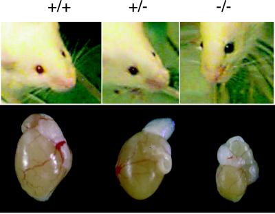
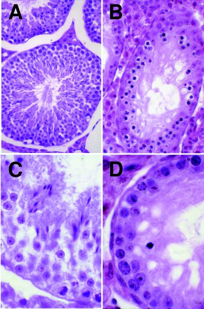
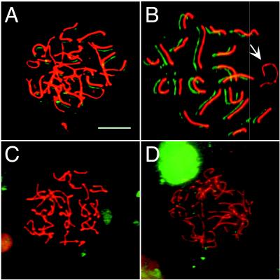
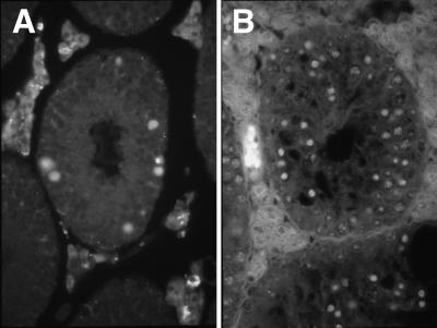
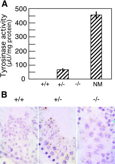
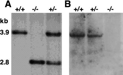
The microrchidia, or morc, autosomal recessive mutation results in the arrest of spermatogenesis early in prophase I of meiosis. The morc mutation arose spontaneously during the development of a mouse strain transgenic for a tyrosinase cDNA construct. Morc -/- males are infertile and have grossly reduced testicular mass, whereas -/- females are normal, indicating that the Morc gene acts specifically during male gametogenesis. Immunofluorescence to synaptonemal complex antigens demonstrated that -/- male germ cells enter meiosis but fail to progress beyond zygotene or leptotene stage. An apoptosis assay revealed massive numbers of cells undergoing apoptosis in testes of -/- mice. No other abnormal phenotype was observed in mutant animals, with the exception of eye pigmentation caused by transgene expression in the retina. Spermatogenesis is normal in +/- males, despite significant transgene expression in germ cells. Genomic analysis of -/- animals indicates the presence of a deletion adjacent to the transgene. Identification of the gene inactivated by the transgene insertion may define a novel biochemical pathway involved in mammalian germ cell development and meiosis.


Figures

Figure 1
Figure 1
Gross appearance of morc mice. (Upper) The wild-type FVB (+/+) pink (albino) eye color contrasts with the brown (+/− mice) or black (−/− mice) eye pigmentation caused by one or two copies of the morc transgene, respectively. (Lower) A marked size reduction is noted in the −/− testis compared with +/+ or +/− testis. Epididymides were of similar size in all three genotypes (the normal epididymis was inadvertently removed from the +/− specimen). The +/− testis is slightly pigmented because of transgene expression (see text).
Figure 2
Figure 2
Testes histology of morc mice. Representative hematoxylin/eosin sections from testes of adult +/− (A and C) or −/− (B and D) mice demonstrate absence of spermatocytes in the −/− animals. Numerous pyknotic cells were present in −/− mice at the location where primary spermatocytes are found. (Magnifications: A and B, ×400; C and D, ×1,000).
Figure 3
Figure 3
Molecular analysis of morc spermatogenic arrest. Immunofluorescence analysis of spermatocytes from 24-day-old morc +/− (A and B) or −/− (C and D) mice, with anti-COR1 (red), recognizing mouse SYCP3, and anti-SYN1 (green), recognizing mouse SYCP1. The green image is deliberately offset from the red image to facilitate visualization of coincidence of staining. (A) Normal zygotene spermatocyte from a +/− mouse, with well-developed axes staining with anti-COR1; these axes are not completely paired, and initiation of synapsis is recognized by staining with anti-SYN1. (B) Normal pachytene spermatocyte from a +/− mouse; note coincident immunoreactivity for SYN1 with all axes indicating full and complete synapsis, except for the sex chromosomes (arrow), which normally synapse only in the most terminal region. (C) Abnormal zygotene-like spermatocyte from a −/− animal. Even though full axial elements are formed, no pairing is seen. (D) A morc −/− zygotene spermatocyte with focal regions of initiation of synapsis. (Bar = 10 μm.)
Figure 4
Figure 4
Apoptosis in testes of adult morc mice. (A) Most (>90%) wild-type tubules revealed no TUNEL positivity; the section shown here demonstrates the occasional apoptotic cells present in wild-type animals. (B) In contrast, the majority of germ cells from −/− mice are undergoing apoptosis. (Magnification: ×200.)
Figure 5
Figure 5
Expression of tyrosinase in morc mice. (A) Tyrosinase activity in testes extracts from morc +/+, +/−, or −/−, as compared with cultured mouse melanocytes (NM). Histograms represent average of triplicate measurements, and bars indicate SD of the mean. (B) Immunohistochemical staining of tyrosinase in testes of morc mice. Wild-type controls (+/+) show no antibody staining. Intense staining is seen in the germ cell cytoplasm of +/− testes. Faint staining is also present in −/− germ cells. (Magnification: ×400.)
Figure 6
Figure 6
Southern blotting using probes flanking the transgene. DNA from +/+, +/−, or −/− animals was digested with EcoRI and hybridized with each end of a 3.9-kb EcoRI fragment containing the transgene insertion site (see text). (A) One end detects an altered fragment (2.8 kb) for the morc chromosome. (B) The opposite end detects the 3.9-kb wild-type fragment with half intensity in +/− animals and no fragment in −/− mice, demonstrating a deletion to one side of the transgene.

References

    1. Sassone-Corsi P. Cell. 1997;88:163–166. - PubMed
    1. Handel M A. In: Spermatogenesis Genetic Aspects. Henning W, editor. Vol. 15. Berlin: Springer; 1987. pp. 1–52.
    1. Vogelweid C M, Verina T, Norton J, Harruff R, Ghetti B. J Neurogenet. 1993;9:89–104. - PubMed
    1. Nantel F, Monaco L, Foulkes N S, Masquilier D, LeMeur M, Henriksen K, Dierich A, Parvinen M, Sassone-Corsi P. Nature (London) 1996;380:159–162. - PubMed
    1. Blendy J A, Kaestner K H, Weinbauer G F, Nieschlag E, Schutz G. Nature (London) 1996;380:162–165. - PubMed

Publication types

MeSH terms

Associated data