. 1998 Nov 24;95(24):14379-83.
doi: 10.1073/pnas.95.24.14379.

Uncoupling signal transducers from oncogenic MET mutants abrogates cell transformation and inhibits invasive growth

Affiliations

Uncoupling signal transducers from oncogenic MET mutants abrogates cell transformation and inhibits invasive growth

A Bardelli et al. Proc Natl Acad Sci U S A. .

Abstract

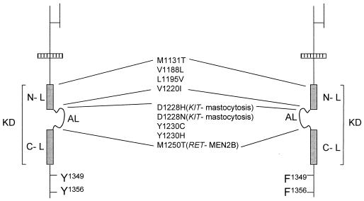
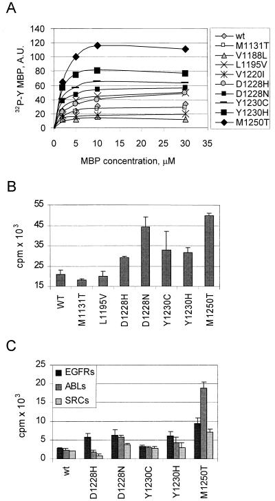
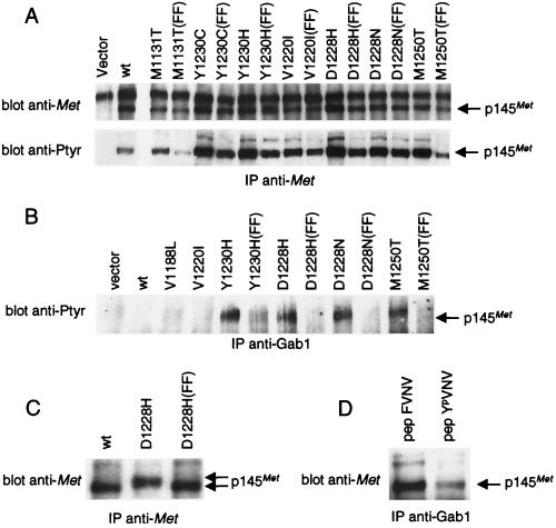
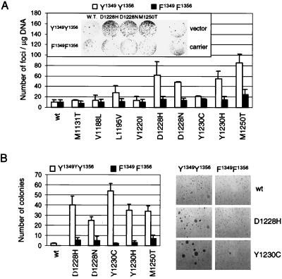
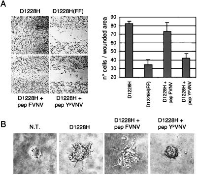
The assumption that genes encoding tyrosine kinase receptors could play a role in human cancers has been confirmed by the identification of oncogenic mutations in the kinase domain of RET and KIT. Recently, homologous residues were found mutated in MET, in papillary renal carcinomas (PRCs). The link coupling these genetic lesions to cellular transformation is still unclear. METPRC mutations result in increased kinase activity and-in some instances, i.e., M1250T substitution-in changes in substrate specificity. A direct correlation occurs between the transforming potential of METPRC mutants and their ability to constitutively associate with signal transducers through two phosphorylated tyrosines (Y1349VHVNATY1356VNV) located in the receptor tail. Substitution of these "docking tyrosines" with phenylalanines leaves unaffected the altered properties of the kinase but abrogates transformation and invasiveness in vitro. Uncoupling the receptor from signal transducers with a tyrosine-phosphorylated peptide derivative (YpVNV) inhibits invasive growth induced by METPRC mutants. These data indicate that constitutive receptor coupling to downstream signal transducers is a key mechanism in neoplastic transformation driven by mutated MET and suggest a therapeutic strategy to target neoplastic diseases associated with this oncogene.


Figures

Figure 1
Figure 1
Map of MET mutations found in PRCs. Schematic representation of the functional domains of the MET tyrosine kinase receptor. Mutations found in PRCs are listed. The tyrosine kinase domain (KD) is represented by a gray box. Y1349 and Y1356, on phosphorylation, generate the docking sites for signal transducers. These tyrosines were converted into phenylalanines (F) to generate PRC mutants unable to bind signal transducers (Right). Mutations of homologous residues found in RET and KIT together with the associated neoplastic diseases are listed in parentheses. AL, activation loop; N-L and C-L, N- and C-terminal kinase lobes.
Figure 2
Figure 2
METPRC mutations affect the kinase properties. Wild-type or PRC-mutated MET receptors were transiently expressed in COS cells, immunoprecipitated, and used to phosphorylate different substrates, including (A) the exogenous substrate myelin basic protein (MBP, (B) the peptide containing the sequence Y1349VHVNATY1356VNV, corresponding to the signal transducer docking site located in the MET tail, and (C) the optimal peptide substrates for the epidermal growth factor transmembrane receptor (EGFRs: AEEEEYFELVAKKKK), the Abl cytosolic kinase (ABLs: EAIYAAPFAKKK), or the Src kinase (SRCs: AEEEIYGEFEAKKK). A shows the amount of 32P incorporated into MBP at different substrate concentrations under the conditions described in Methods. Symbols indicate the METPRC proteins harboring the mutation listed on the right. B and C show the extent of phosphorylation of the indicated peptide by METPRC mutants.
Figure 3
Figure 3
METPRC mutants are constitutively coupled to signal transducers via two tyrosines located in the tail of the receptor. (A) NIH 3T3 fibroblasts were transfected with either wild-type or PRC-mutated MET receptor mutants to generate stable cell lines. FF indicates MET-receptor variants in which tyrosines Y1349 and Y1356 were converted into phenylalanines. The amount of Met proteins and the level of phosphorylation was evaluated by immunoprecipitation with anti-Met antibodies followed by Western blotting with anti-Met or anti-phosphotyrosine (pTyr) antibodies, respectively. (B) Lysates of the same cells were immunoprecipitated with anti-Gab1 antibodies and analyzed by Western immunoblotting with anti-pTyr antibodies. The identity of the phosphorylated protein coimmunoprecipitated with Gab1 was confirmed by reprobing the same blot with an anti-Met antibody (data not shown). (C) Lysates of COS cells expressing either the wild-type (wt), the D1228H mutant, or its double phenylalanine counterpart D1228H (FF) were immunoprecipitated and analyzed by Western blotting with anti-Met antibodies. The gap between the arrows indicates the electrophoretic shift caused by differences in phosphorylation. (D) Lysates of NIH 3T3 cells expressing the D1228H mutant were incubated with the YpVNV phosphopeptide corresponding to the receptor’s docking site and immunoprecipitated with anti-Gab1 antibodies. The amount of associated receptor was determined by Western blot with anti-Met antibodies. The nonphosphorylated phenylalanine peptide analogue (FVNV) was used as a negative control. The arrows indicate the position of the 145-kDa MET receptor β-chain (p145Met).
Figure 4
Figure 4
Uncoupling METPRC mutants from signal transducers abrogates transformation and anchorage-independent growth. (A) The transforming potential of METPRC mutants (□, Y1349–Y1356) and of their signaling-inactive counterparts (■, F1349–F1356) was evaluated using the focus-forming assay. Values reported represent the average of three independent experiments (±SD). (Inset) micrograph showing a representative plate for each MET mutant after 3 weeks. Vector and carrier indicate cells transfected with empty vector or with calf-thymus DNA, respectively. (B) Anchorage-independent growth of METPRC mutant (□, Y1349–Y1356) and of their signaling-inactive counterparts (■, F1349–F1356) was assessed in soft agar. Values represent the average of two independent experiments (±SD). Micrographs show representative fields of colonies after 3 weeks of growth in soft agar.
Figure 5
Figure 5
Uncoupling METPRC mutants from signal transducers inhibits invasive growth. (A) The invasive potential of the D1228H mutants and of its signaling-inactive counterpart D1228H (FF) was evaluated by measuring the ability to invade reconstituted basal membranes made of laminin, collagen type IV, and proteoglycans (Matrigel). The YpVNV phosphopeptide was introduced into living cells by in situ electroporation; monolayers were then wounded and covered with Matrigel. The nonphosphorylated peptide FVNV was used as a negative control. The invasive phenotype was quantified by measuring the number of cells (±SD) found in the wounded area 12 hr later (see Methods). Micrographs show representative fields of cells plated on conductive slides used for in situ electroporation. (B) The invasive growth of the D1228H mutant was evaluated by measuring the sprouting and migration into a three-dimensional network of collagen type I. Human kidney cells (BOSC-23) either nontransfected (N.T.) or transiently expressing the D1228H receptor were seeded in collagen gels and grown for 5 days. The indicated peptides were introduced by in situ cell electroporation. Micrographs of representative fields were taken after 5 days.

Similar articles

Cited by

References

    1. Mulligan L M, Kwok J B, Healey C S, Elsdon M J, Eng C, Gardner E, Love D R, Mole S E, Moore J K, Papi L, et al. Nature (London) 1993;363:458–460. - PubMed
    1. Hofstra R M, Landsvater R M, Ceccherini I, Stulp R P, Stelwagen T, Luo Y, Pasini B, Hoppener J W, van Amstel H K, Romeo G, et al. Nature (London) 1994;367:375–376. - PubMed
    1. Nagata H, Worobec A S, Oh C K, Chowdhury B A, Tannenbaum S, Suzuki Y, Metcalfe D D. Proc Natl Acad Sci USA. 1995;92:10560–10564. - PMC - PubMed
    1. Longley B J, Tyrrell L, Lu S Z, Ma Y S, Langley K, Ding T G, Duffy T, Jacobs P, Tang L H, Modlin I. Nat Genet. 1996;12:312–314. - PubMed
    1. Hirota S, Isozaki K, Moriyama Y, Hashimoto K, Nishida T, Ishiguro S, Kawano K, Hanada M, Kurata A, Takeda M, et al. Science. 1998;279:577–580. - PubMed

Publication types

MeSH terms