. 1998 Nov 24;95(24):14389-94.
doi: 10.1073/pnas.95.24.14389.

Vascular endothelial growth factor C induces angiogenesis in vivo

Affiliations

Vascular endothelial growth factor C induces angiogenesis in vivo

Y Cao et al. Proc Natl Acad Sci U S A. .

Abstract

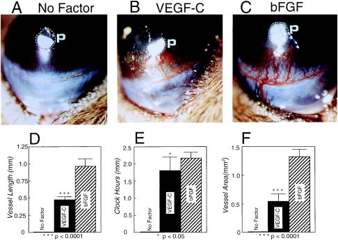
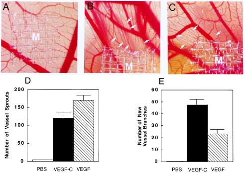
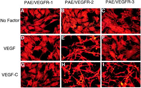
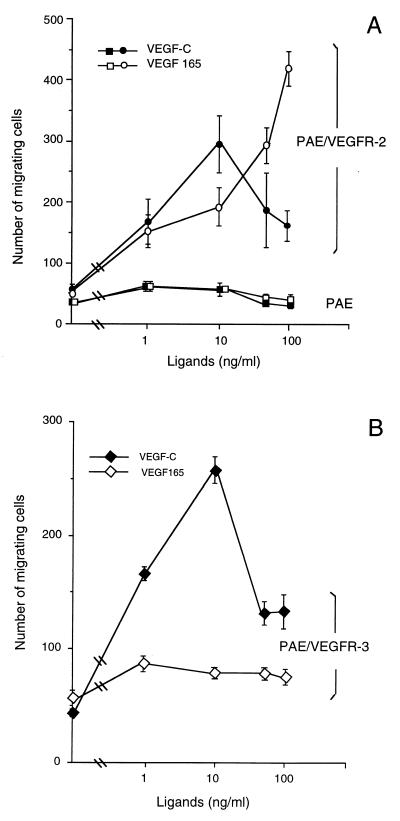
Vascular endothelial growth factor C (VEGF-C) recently has been described to be a relatively specific growth factor for the lymphatic vascular system. Here we report that ectopic application of recombinant VEGF-C also has potent angiogenic effects in vivo. VEGF-C is sufficiently potent to stimulate neovascularization from limbal vessels in the mouse cornea. Similar to VEGF, the angiogenic response of corneas induced by VEGF-C is intensive, with a high density of new capillaries. However, the outgrowth of microvessels stimulated by VEGF-C was significantly longer than that induced by VEGF. In the developing embryo, VEGF-C was able to induce branch sprouts from the established blood vessels. VEGF-C also induced an elongated, spindle-like cell shape change and actin reorganization in both VEGF receptor (VEGFR)-2 and VEGFR-3-overexpressing endothelial cells, but not in VEGFR-1-expressing cells. Further, both VEGFR-2 and VEGFR-3 could mediate proliferative and chemotactic responses in endothelial cells on VEGF-C stimulation. Thus, VEGF-C may regulate physiological angiogenesis and participate in the development and progression of angiogenic diseases in addition to lymphangiogenesis.


Figures

Figure 1
Figure 1
SDS/PAGE analysis of VEGF-C preparation. Recombinant mature form of human VEGF-C was expressed in pichia pastoris yeast cells. Three micrograms of the purified VEGF-C was analyzed on a 10–20% gradient SDS-polyacrylamide gel followed by staining with Coomassie blue. Dimeric (lane 2) and monomeric (lane 3) forms of VEGF-C were detected under nonreducing (lane 2) and reducing/alkylating conditions (lane 3). Molecular mass markers are indicated on the left (lane 1).
Figure 2
Figure 2
Corneal neovascularization stimulated by VEGF-C and VEGF. Pellets of sucrose aluminum sulfate and hydron polymer containing 160 ng of VEGF-C (A) and VEGF (B) were implanted into corneal micropockets of C57BL6/J mice as described in Materials and Methods. Corneas were photographed with a slit-lamp stereomicroscope on day 5 after growth factor implantation. Dashed lines encircle the area of the implanted pellets. Photographs represent ×20 amplification of the mouse eye. Quantitation of corneal neovascularization. (C) Maximal vessel length. (D) Clock hours of circumferential neovascularization. (E) Area of neovascularization. Graphs represent mean values (±SEM) of 10 eyes (five mice) in each group. The P values were calculated by two-tailed Student t test analysis using the statistical instat 1.1 and Microsoft excel 5 programs.
Figure 3
Figure 3
Angiogenic effects of VEGF-C and FGF-2 in the cornea. Pellets containing 160 ng of VEGF-C (B), 80 ng of FGF-2 (C), and PBS alone without growth factors (A) were implanted into corneal micropockets of C57BL6/J mice. Photographs represent ×20 amplification of the mouse eye. p, pellets. Corneal neovascularization was quantitated on day 5 after pellet implantation. (D) Maximal vessel length. (E) Clock hours of circumferential neovascularization. (F) Area of neovascularization. Graphs represent mean values (±SEM) of 10 eyes of five mice in each group.
Figure 4
Figure 4
Angiogenic effect of VEGF-C and VEGF on CAMs. Nylon meshes (9.3 mm2) coated with methylcellulose containing 2.5 μg of VEGF-C and VEGF were implanted on CAMs of 7-day-old chicken embryos. After 4-day implantation, the formation of new blood vessels was examined under a stereoscope. (A) A control CAM with a methylcellulose mesh containing PBS without growth factors. An example of VEGF-C- (B) and VEGF-implanted CAM (C). New blood vessel branches are marked by large arrows. Small arrows indicate the new microvessel sprouts. At the concentration of 2.5 μg/disk, the number of vessel sprouts (D) and the number of new vessel branches (E) stimulated by VEGF-C, VEGF, and PBS were quantitated within a defined area of 85 mm2 surrounding the implanted mesh. Seven embryos in each group were used for quantitative studies. Data represent mean values (±SEM).
Figure 5
Figure 5
VEGF-C- and VEGF-induced endothelial cell shape changes and actin reorganization. VEGF-C and VEGF were added to approximately 70% confluent VEGF receptor-expressing PAE cells at a final concentration of 100 ng/ml for 12 h, fixed with paraformaldehyde, permeabilized with Triton X-100, and stained with rhodamine-phalloidin. In response to VEGF-C (GI) and VEGF (DF), PAE/VEGFR-2 cells (H and E) undergo dramatic spindle-like shape changes, leading lamellae, lamellipodium extensions and actin reorganization. PAE/VEGFR-3 cells also show this striking cell shape change in response to VEGF-C stimulation (I). PAE/VEGFR-1 cells lack such a cell shape change in the presence of VEGF-C (G) and VEGF (D). All VEGFR-expressing cells also lack cell morphological changes in the absence of growth factors (AC).
Figure 6
Figure 6
Cell proliferation. The proliferative effects of VEGF-C (■) and VEGF (○) were assayed on PAE/VEGFR-1 (A), PAE/VEGFR-2 (B), and PAE/VEGFR-3 (C) cells. VEGF-C and VEGF at various concentrations in a triplicate of each sample were incubated with cells in the presence of 1% FCS. After 72 h incubation, cell numbers were counted by a Coulter counter, and values represent the mean (±SEM) of a triplicate of each sample. PBS was used as the negative control.
Figure 7
Figure 7
Chemotactic activity. (A) Chemotactic response of VEGFR-2/PAE cells (circles) and nontransfected PAE cells (squares) to VEGF (open symbols) and VEGF-C and (filled symbols). (B) Chemotactic response of VEGFR-3 cells to VEGF-C (○) and VEGF (■). Cells were incubated in serum-free Ham′s F-12 medium containing 0.2% BSA with or without growth factors for 4 h at 37°C in a modified Boyden chamber. Migrating cells were counted after staining with Giemsa and plotted in absolute number per optic field. Data represent means (±SEM) of a triplicate of each sample.

Similar articles

Cited by

References

    1. Folkman J, Shing Y. J Biol Chem. 1992;267:10931–10934. - PubMed
    1. Folkman J. Nat Med. 1995;1:27–31. - PubMed
    1. Hanahan D, Folkman J. Cell. 1996;86:353–364. - PubMed
    1. Folkman J. N Engl J Med. 1995;333:1757–1763. - PubMed
    1. Senger D R, Galli S J, Dvorak A M, Perruzzi C A, Harvey V S, Dvorak H F. Science. 1983;219:983–985. - PubMed

Publication types

MeSH terms

Substances

LinkOut - more resources