. 1998 Nov 24;95(24):14558-63.
doi: 10.1073/pnas.95.24.14558.

Rat heart: a site of oxytocin production and action

Affiliations

Rat heart: a site of oxytocin production and action

M Jankowski et al. Proc Natl Acad Sci U S A. .

Abstract

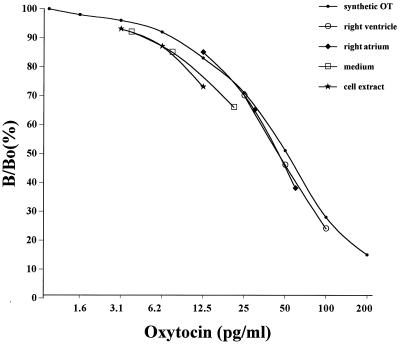
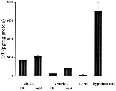
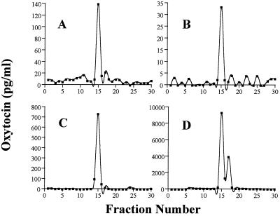
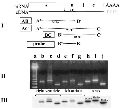
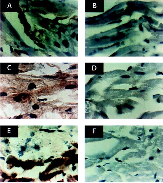
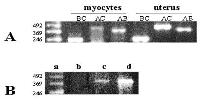
We report here that the rat heart is a site of oxytocin (OT) synthesis and release. Oxytocin was detected in all four chambers of the heart. The highest OT concentration was in the right atrium (2128 +/- 114 pg/mg protein), which was 19-fold higher than in rat uterus but 3.3-fold lower than in the hypothalamus. OT concentrations were significantly greater in the right and left atria than in the corresponding ventricles. Furthermore, OT was released into the effluent of isolated, perfused rat heart (34.5 +/- 4.7 pg/min) and into the medium of cultured atrial myocytes. Reverse-phase HPLC purification of the heart extracts and heart perfusates revealed a main peak identical with the retention time of synthetic OT. Southern blots of reverse transcription-PCR products from rat heart revealed gene expression of specific OT mRNA. OT immunostaining likewise was found in atrial myocytes and fibroblasts, and the intensity of positive stains from OT receptors paralleled the atrial natriuretic peptide stores. Our findings suggest that heart OT is structurally identical, and therefore derived from, the same gene as the OT that is primarily found in the hypothalamus. Thus, the heart synthesizes and processes a biologically active form of OT. The presence of OT and OT receptor in all of the heart's chambers suggests an autocrine and/or paracrine role for the peptide. Our finding of abundant OT receptor in atrial myocytes supports our hypothesis that OT, directly and/or via atrial natriuretic peptide release, can regulate the force of cardiac contraction.


Figures

Figure 1
Figure 1
Oxytocin immunoreactivity of serial dilutions (1:80; 1:40; 1:20) of rat extracts from atrial and ventricular homogenates as well as medium and cell extracts from cultures of atrial myocytes in vitro were measured by radioimmunoassay. Results are compared with the oxytocin standard curve. The ordinate shows the ratio (expressed as the percentage) of bound 125I-labeled oxytocin in the presence (B) and in the absence (Bo) of synthetic oxytocin.
Figure 2
Figure 2
Oxytocin concentration in rat tissues obtained by RIA after prior extraction by Vycor heat-activated glass beads.
Figure 3
Figure 3
Reverse-phase HPLC elution profile of oxytocin immunoreactivity in rat tissues. Tissue extracts, lyophilized and reconstituted in 20% acetonitrile in 0.1% TFA, were applied to a C18 Bondapak column and were eluted with acetonitrile gradient (20–50%) in 0.1% TFA. (A) The extract from the right atrium. (B) The extract from the heart perfusates. (C) The HPLC profile of OT synthetic standard. (D) The extract from the pituitary gland.
Figure 4
Figure 4
RT and PCR analysis of rat cardiac OT mRNA based on the concept presented by Lefebvre et al. (17). (I) Schematic representation of the approximate positions of the primers used in the RT-PCR amplification and the probe used to determine the specificity of the PCR products. A+, exon A-specific primer sense-strand, primer corresponding to sequences +3 and +26 OT gene; B+, exon B-specific sense-strand primer (+416 to + 434); B−, exon B-specific antisense-strand primer (+559 to +584); C−, exon C-specific antisense-strand primer (+838 to +864). (II) Ethidium bromide-stained agarose gel showing the PCR amplification products obtained with OT exon-specific primers by using reverse-transcribed mRNA from rat right ventricle, left atrium, and uterus, used as control organ. Lanes: a, 123-bp ladder of GIBCO/BRL; b, e, and h, AB OT cDNA products of PCR; c, f, and I, AC OT cDNA products of PCR; d, g, and j, BC OT cDNA products of PCR. (III) Southern blot analysis of PCR amplification products resolved in an agarose gel, transferred to nylon membrane, and hybridized with B+B− OT cDNA probe.
Figure 5
Figure 5
Immunocytochemical staining of sections of rat atrium (A) and ventricle (B) with rabbit anti-OT antibody at final dilution of 1/250 and atrium (C) and ventricle (D) with rabbit anti-OTR antibody diluted 1/250. The results are related to control sections cut on the border of the atrium (lower part of images) and the ventricle (upper part of images) reacted with anti-ANP rabbit antibody diluted 1/500 (E) and normal rabbit serum (F). The specific immunostaining is visible as a yellow-brownish color after incubation with F(ab′)2 fragments of horseradish peroxidase-conjugated goat anti-rabbit IgG and diaminobenzidine. The sections were counterstained with hematoxylin.
Figure 6
Figure 6
Detection of oxytocin mRNA and oxytocin receptor mRNA in rat atrial myocytes by RT-PCR. (A) Amplification of cDNA with OT-exon-specific primers (as shown in Fig. 4). The photograph presents RT-PCR products after electrophoresis on 2% agarose in ethidium bromide. Lanes: a, 123-bp ladder GIBCO/BRL; b, c, and d, OT-exon-specific PCR product amplified from the cDNA of atrial myocytes; e, f, and g, the corresponding PCR OT gene products amplified from uterine cDNA. (B) OTR RT-PCR products after electrophoresis on 2% agarose. Lanes, a, 123-bp ladder GIBCO/BRL; b, not reverse-transcribed total RNA from atrial myocytes; c, atrial myocytes; d, uterus.

References

    1. Swanson L W, Kuypers H G J M. J Comp Neurol. 1980;194:555–570. - PubMed
    1. Heller H. In: Occurrence, Storage and Metabolism of Oxytocin. Caldeyro-Barcia R, Heller H, editors. New York: Pergamon; 1961. pp. 3–23.
    1. Chalmers J, Pilowsky P. J Hypertens. 1991;9:675–694. - PubMed
    1. Jirikowski G F, Back H, Forssmann W G, Stumpf W E. Neuropeptides. 1986;8:243–249. - PubMed
    1. Petersson M, Alster P, Lundeberg T, Uvnäs-Moberg K S. Physiol Behav. 1996;60:1311–1315. - PubMed

Publication types

MeSH terms

LinkOut - more resources