Review
. 1998 Nov;21(11):471-6.
doi: 10.1016/s0166-2236(98)01275-2.

Neuropeptide-mediated excitability: a key triggering mechanism for seizure generation in the developing brain

Affiliations
Review

Neuropeptide-mediated excitability: a key triggering mechanism for seizure generation in the developing brain

T Z Baram et al. Trends Neurosci. 1998 Nov.

Abstract

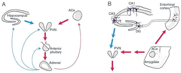
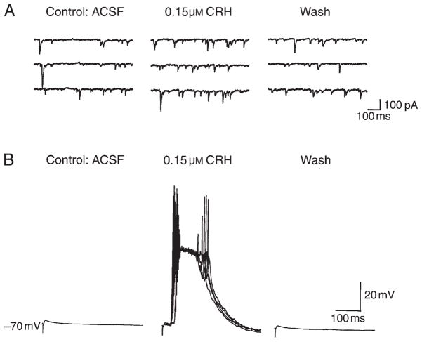
Most human seizures occur early in life,consistent with established excitability-promoting features of the developing brain. Surprisingly, the majority of developmental seizures are not spontaneous but are provoked by injurious or stressful stimuli. What mechanisms mediate'triggering' of seizures and limit such reactive seizures to early postnatal life? Recent evidence implicates the excitatory neuropeptide, corticotropin-releasing hormone (CRH). Stress activates expression of the CRH gene in several limbic regions, and CRH-expressing neurons are strategically localized in the immature rat hippocampus, in which this neuropeptide increases the excitability of pyramidal cells in vitro. Indeed, in vivo, activation of CRH receptors--maximally expressed in hippocampus and amygdala during the developmental period which is characterized by peak susceptibility to 'provoked' convulsions--induces severe, age-dependent seizures. Thus, converging data indicate that activation of expression of CRH constitutes an important mechanism for generating developmentally regulated, triggered seizures, with considerable clinical relevance.


Figures

Fig. 1
Fig. 1. The neuroendocrine (A) and limbic (B) interactive, stress-activated corticotropin-releasing hormone (CRH) loops
(A) Stress-conveying signals rapidly activate immediate–early genes in CRH-expressing neurons of the central nucleus of the amygdala (ACe). Rapid CRH release in the ACe is associated with CRH-expression in hypothalamic paraventricular nucleus (PVN) and secretion of CRH into the hypothalamo–pituitary portal system, inducing ACTH and glucocorticoid secretion from the pituitary and adrenal, respectively. Glucocorticoids exert a negative feedback on the PVN (directly and via the hippocampus), yet activate expression of the CRH gene in the amygdala, potentially promoting further CRH release in this region. (B) CRH-expressing GABAergic interneurons (purple cells) in the principal cell layers of the hippocampal CA1, CA3 and the dentate gyrus (DG) are positioned to control excitability of the pyramidal and granule cells, respectively. These neurons might be influenced by stress-evoked release of CRH from the ACe, via connections in the entorhinal cortex. For both panels, red and blue arrows denote established or putative potentiating and inhibitory actions, respectively. Arrows do not imply monosynaptic connections.
Fig. 2
Fig. 2. Rapid activation of expression of the corticotropin-releasing hormone (CRH) gene by stress in the hypothalamic paraventricular nucleus
Coronal brain sections were subjected to in situ hybridization using an intronic probe recognizing only heteronuclear RNA (hnRNA, unedited) encoding CRH. (A) Sections derived from stress-free 9-day old pups, sacrificed within 20 s of disturbance, show little CRH hnRNA-specific signal. (B) CRH expression is evident, by 2 min from the onset of stress, and (C) peaks by 15 min. CRH hnRNA levels decline by 30 min from the onset of stress (D), while total CRH-specific mRNA is increased for at least 4 h after stress. Scale bar in (A), 300 μm.
Fig. 3
Fig. 3. Distribution and characterization of corticotropin-releasing hormone (CRH)-expressing neurons in the hippocampus
(A) Immunolabeled CRH-expressing neurons within and adjacent to the pyramidal (s.p.) layer of CA3. (B) A double-labeled neuron, expressing both CRH (brown) and an isoform of glutamic acid decarboxylase (GAD-65), the GABA-synthesizing enzyme (blue–green). (C) An interneuron reacting with antibodies to both CRH (orange) and parvalbumin (plum red). Scale bar, 50 μm in (A), 15 μm in (B) and 20 μm in (C). Abbreviations: s.o., stratum oriens; s.r., stratum radiatum. Modified, with permission, from Yan et al.
Fig. 4
Fig. 4. Corticotropin-releasing hormone (CRH) enhances excitability of hippocampal pyramidal neurons
(A) Reversible increase in the frequency of spontaneous excitatory postsynaptic currents with application of CRH (middle panel). Whole-cell recording of a CA3 pyramidal neuron was performed in voltage-clamp mode (−60 mV). (B) Large, synchronized, variable-latency discharges of CA3 neurons in current-clamp mode (in the presence of bicuculline) are elicited by stimulation of mossy fibers in the presence of CRH. These action-potential discharges are longer-latency, probably polysynaptic, excitatory postsynaptic potentials, and are fully reversed when CRH is removed. Abbreviation: ACSF, artificial cerebrospinal fluid. Modified, with permission, from Hollrigel et al.

References

    1. Berg AT, Shinnar S. J Child Neurol. 1994;9 (Suppl 2):S19–S26. - PubMed
    1. Berg AT, et al. Epilepsia. 1995;36:334–341. - PubMed
    1. Baram TZ, Gerth A, Schultz L. Dev Brain Res. 1997;246:134–143. - PMC - PubMed
    1. Holtzman D, Obana K, Olson J. Science. 1981;213:1034–1036. - PubMed
    1. Jensen FE, et al. Ann Neurol. 1991;29:629–637. - PubMed

Publication types