. 1998 Dec;180(23):6224-31.
doi: 10.1128/JB.180.23.6224-6231.1998.

Identification and characterization of SpcU, a chaperone required for efficient secretion of the ExoU cytotoxin

Affiliations

Identification and characterization of SpcU, a chaperone required for efficient secretion of the ExoU cytotoxin

V Finck-Barbançon et al. J Bacteriol. 1998 Dec.

Abstract

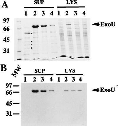
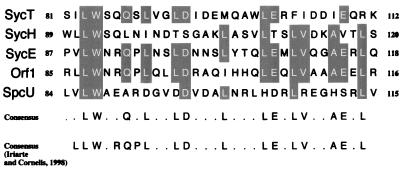
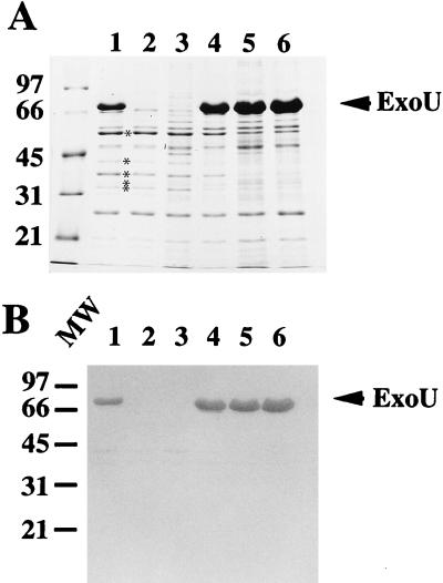
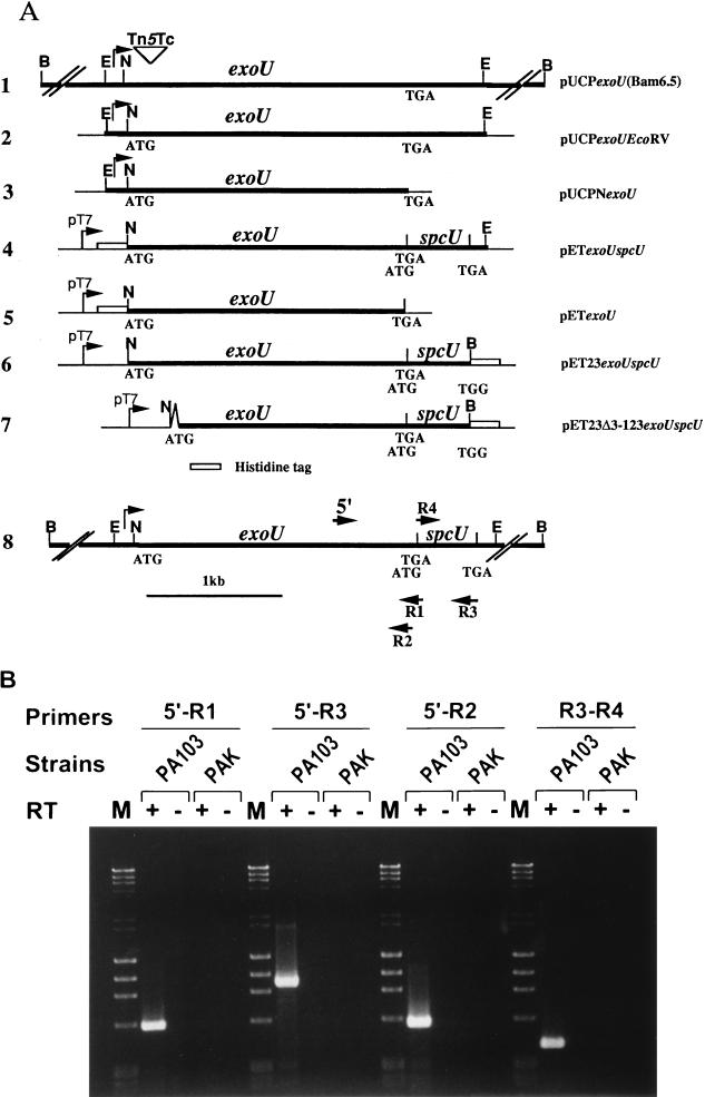
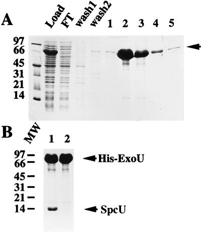
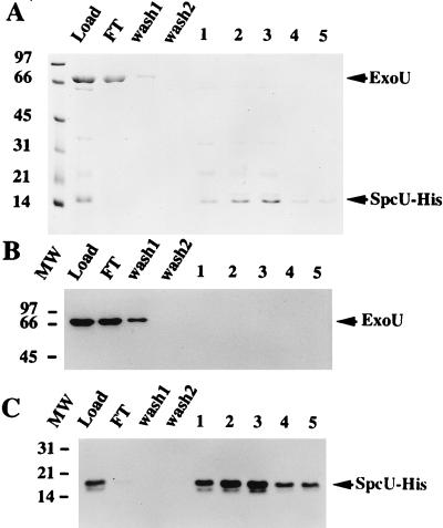
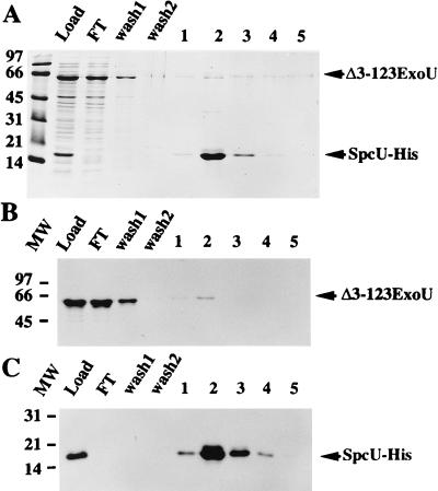
In recent studies, we have shown that Pseudomonas aeruginosa strains that are acutely cytotoxic in vitro damage the lung epithelium in vivo. Genetic analysis indicated that the factor responsible for acute cytotoxicity was controlled by ExsA and therefore was part of the exoenzyme S regulon. The specific virulence determinant responsible for epithelial damage in vivo and cytotoxicity in vitro was subsequently mapped to the exoU locus. The present studies are focused on a genetic characterization of the exoU locus. Northern blot analyses and complementation experiments indicated that a region downstream of exoU was expressed and that the expression of this region corresponded to increased ExoU secretion. DNA sequence analysis of a region downstream of exoU identified several potential coding regions. One of these open reading frames, SpcU (specific Pseudomonas chaperone for ExoU), encoded a small 15-kDa acidic protein (137 amino acids [pI 4.4]) that possessed a leucine-rich motif associated with the Syc family of cytosolic chaperones for the Yersinia Yops. T7 expression analysis and nickel chromatography of histidine-tagged proteins indicated that ExoU and SpcU associated as a noncovalent complex when coexpressed in Escherichia coli. The association of ExoU and SpcU required amino acids 3 to 123 of ExoU. In P. aeruginosa, ExoU and SpcU are coordinately expressed as an operon that is controlled at the transcriptional level by ExsA.


Figures

FIG. 1
FIG. 1
Complementation of PA103exoU::Tn5Tc. (A) SDS-polyacrylamide gel (11% acrylamide), stained with Coomassie blue R250, of protein profiles of P. aeruginosa PA103exoU::Tn5Tc (a polar insertion within exoU, host strain) containing different plasmid constructs of the ExoU locus. All strains were grown under inducing conditions for expression of the exoenzyme S regulon. Equivalent amounts (normalized to the culture optical density at 540 nm) of either extracellular fractions (supernatant [SUP]) or lysate (LYS) preparations were loaded in each lane. Lanes: 1, P. aeruginosa PA103exoU::Tn5Tc; 2, PA103exoU::Tn5Tc complemented in trans with pUCPexoU(Bam 6.5); 3, the host strain with pUCPexoUEcoRV; and 4, the host strain with pUCPNexoU. The positions of other type III-secreted products of the exoenzyme S regulon are shown by asterisks in panel A (in descending order, ExoT, PopB, PopD, PopN, and PcrV). (B) Western Blot analysis of a duplicate gel probed with polyclonal anti-ExoU antibodies. The positions of ExoU (arrow) and molecular weight (MW) standards (left [thousands]) are indicated.
FIG. 2
FIG. 2
Pileup alignment of the leucine repeat motif from Syc and Syc-like chaperones. Amino acids 84 to 115 of SpcU were aligned to amino acids 85 to 116 of Orf1 (putative chaperone for ExoS or ExoT) (41), amino acids 87 to 118 of SycE (37), amino acids 89 to 120 of SycH (35), and amino acids 81 to 112 of SycT (18). Common amino acids that conform to the consensus sequence are presented in shaded boxes. A consensus sequence, which includes SpcU as part of the alignment, is shown. A consensus sequence, as determined by Iriarte and Cornelis (18), which includes a leucine-rich motif, is indicated for comparison.
FIG. 3
FIG. 3
Complementation of PA103ΔexoU. (A) SDS-PAGE analysis (11% acrylamide; Coomassie-stained gel) of equivalent amounts of extracellular proteins from P. aeruginosa strains grown under inducing conditions for the exoenzyme S regulon. Lanes: 1, strain PA103; 2, PA103ΔexoU (a nonpolar deletion in exoU); 3, PA103ΔexoU containing the vector control plasmid pUCP19; 4, PA103ΔexoUpUCPNexoU; 5, PA103ΔexoUpUCPexoUEcoRV; 6, PA103ΔexoUpUCPexoU(Bam 6.5). The positions of other type III-secreted products of the exoenzyme S regulon are shown by asterisks in panel A (in descending order, ExoT, PopB, PopD, PopN, and PcrV). (B) Western blot of a duplicate gel probed with polyclonal anti-ExoU antibodies. The positions of ExoU (arrow) and molecular weight (MW) standards (left [thousands]) are indicated.
FIG. 4
FIG. 4
ExoU locus constructs and amplification of the exoU-spcU transcript by RT-PCR. (A) Schematic representation of the location of the exoU and spcU ORFs within different plasmid constructs used in complementation analyses in P. aeruginosa (constructs, lines 1 to 3), protein expression and purification in E. coli (constructs, lines 4 to 7), and mapping of the mRNA 3′ end (line 8). Double slash marks indicate regions that are not drawn to scale. The chromosomal insertion of the polar Tn5 insertion within the 5′ coding region of exoU is illustrated by an inverted triangle (line 1). Corresponding constructs include pUCPexoU(Bam6.5) (line 1), pUCPexoUEcoRV (line 2), pUCPNexoU (line 3), pETexoUspcU (line 4), pETexoU (line 5), pET23exoUspcU (line 6), and pET23Δ3–123exoUspcU (line 7). N, NsiI; E, EcoRV; B, BamHI. An inverted V in the construct on line 7 represents the deletion of amino acids 3 to 123 of ExoU. Line 8, map of the ExoU locus with the approximate locations of primers used for PCR amplification. Total RNAs from P. aeruginosa strains PA103 (exoU+) and PAK (exoU) were used for the first-strand cDNA synthesis in the presence (+) or absence (−) of Superscript II RT. RT-PCR was performed as described in Materials and Methods. (B) Products of the RT-PCRs were analyzed on a 1.2% agarose gel. PA103 (exoU+) and PAK (exoU) strains are indicated over the brackets, and the primer pairs used for each reaction are underlined. M, molecular weight markers. + and −, reaction mixtures containing (+) or not containing (−) RT.
FIG. 5
FIG. 5
Nickel chromatography, under native conditions, of recombinant amino-terminally-tagged ExoU. (A) SDS-PAGE (13.5% polyacrylamide stained with Coomassie blue R250) analysis of the cell lysate from host strain E. coli BL21(DE3) pLysS with pETexoU (N-terminal 10-histidine tag). Protein profiles of the cell lysate are shown as the material loaded onto a nickel column (Load), the flowthrough from the column (FT), wash fractions 1 and 2, and the elution fractions 1 to 5 in the presence of high concentrations of imidazole. The arrow denotes the position of His-ExoU. (B) SDS-PAGE analysis of peak eluate fractions from nickel chromatography of two expression constructs. Lane 1 contains the peak eluate fraction from an expression experiment using the pETexoUspcU construct to produce recombinant protein (His-ExoU and SpcU). Lane 2 contains a similar fraction from the expression of pETexoU (His-ExoU). Molecular weight (MW) standards (left [thousands]) and recombinant proteins (arrows) are indicated.
FIG. 6
FIG. 6
Denaturing nickel chromatography of His-ExoU. An N-terminal histidine-tagged ExoU construct also encoding an untagged version of SpcU (pETexoUspcU) was expressed in E. coli BL21(DE3) pLysS. The peak eluate fraction from a chromatography performed under nondenaturing conditions (Fig. 5B, lane 1) was collected in a urea solution (final concentration of 6 M). After dialysis to eliminate imidazole, the denatured sample was loaded onto a nickel column equilibrated in urea. (A) SDS-PAGE analysis (13.5% polyacrylamide) of the column fractions which include the load, flowthrough (FT), wash fractions 1 and 2, and elution fractions (in the presence of a high concentration of imidazole) 1 to 5. (B) Western blot analysis of a duplicate gel probed with polyclonal antiserum specific for ExoU. Bound IgG was detected by 125I-protein A. His-ExoU and SpcU (arrows) and molecular weight (MW) standards (left [thousands]) are indicated.
FIG. 7
FIG. 7
Denaturing nickel chromatography of SpcU-His. A C-terminal six-histidine-tagged SpcU-His fusion construct also encoding an untagged version of ExoU was expressed from E. coli BL21(DE3) pET23exoUspcU. The eluate fractions from a native chromatography of the cell lysate were pooled, dialyzed to eliminate imidazole, brought to 6 M urea, and loaded onto a nickel column equilibrated in urea. (A) SDS-PAGE (13.5% polyacrylamide) analysis of the protein profiles of the different fractions: load, flowthrough (FT), wash fractions (wash 1 and 2), and elution fractions 1 to 5. (B) Western blot analysis of a duplicate gel (as in panel A) probed with polyclonal antiserum specific for ExoU. (C) Western blot analysis of a duplicate gel (as in panel A) probed with a primary monoclonal antibody specific for the histidine tag and a secondary rabbit anti-mouse IgG. Bound IgG was detected in blots B and C by using 125I-protein A. Arrows on the right indicate the migration of ExoU and SpcU-His. The molecular weight (MW) markers are indicated on the left (thousands).
FIG. 8
FIG. 8
Amino acids 3 to 123 of ExoU are required to bind to SpcU-His. The cell lysate from E. coli BL21(DE3)pLysS pET23Δ3–123exoUspcU was subjected to nickel affinity chromatography under native conditions. This construct encodes an amino-terminal deletion derivative of ExoU and SpcU-His. (A) SDS-PAGE analysis (13.5% polyacrylamide) of the protein profiles of the different fractions: load, flowthrough (FT), wash fractions 1 and 2, and elution fractions 1 to 5. (B) Western blot analysis of a duplicate gel (as shown in panel A) probed with polyclonal antiserum specific for ExoU. (C) Western blot analysis of a duplicate gel (as shown in panel A) probed with a primary monoclonal antibody specific for the histidine tag and a secondary rabbit anti-mouse IgG. Bound IgG was detected by 125I-protein A. Arrows on the right indicate the migration of Δ3–123ExoU and SpcU-His. The molecular weight (MW) markers are indicated on the left (thousands).

References

    1. Anderson D, Schneewind O. A mRNA signal for the type III secretion of Yop proteins by Yersinia enterocolitica. Science. 1997;278:1140–1143. - PubMed
    1. Bette-Bobillo P, Giro P, Sainte-Marie J, Vidal M. Exoenzyme S from P. aeruginosa ADP ribosylates rab4 and inhibits transferrin recycling in SLO-permeabilized reticulocytes. Biochem Biophys Res Commun. 1998;244:336–341. - PubMed
    1. Cheng L W, Anderson D M, Schneewind O. Two independent type III secretion mechanisms for YopE in Yersinia enterocolitica. Mol Microbiol. 1997;24:757–765. - PubMed
    1. Cornelis G R, Wolf-Watz H. The Yersinia Yop virulon: a bacterial system for subverting eukaryotic cells. Mol Microbiol. 1997;23:861–867. - PubMed
    1. Finck-Barbançon V, Goranson J, Zhu L, Sawa T, Wiener-Kronish J P, Fleiszig S M J, Wu C, Mende-Mueller L, Frank D W. ExoU expression by Pseudomonas aeruginosa correlates with acute cytotoxicity and epithelial injury. Mol Microbiol. 1997;25:547–557. - PubMed

Publication types

MeSH terms

Associated data