. 1998 Dec;180(23):6232-41.
doi: 10.1128/JB.180.23.6232-6241.1998.

Role of SarA in virulence determinant production and environmental signal transduction in Staphylococcus aureus

Affiliations

Role of SarA in virulence determinant production and environmental signal transduction in Staphylococcus aureus

P F Chan et al. J Bacteriol. 1998 Dec.

Abstract

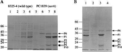
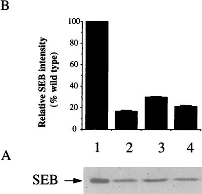
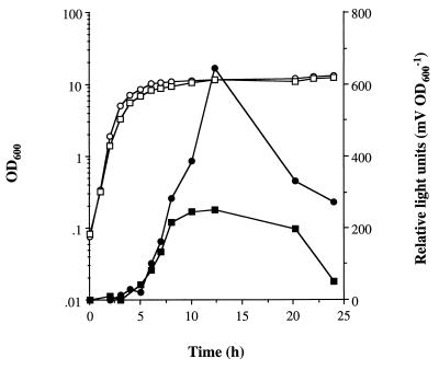
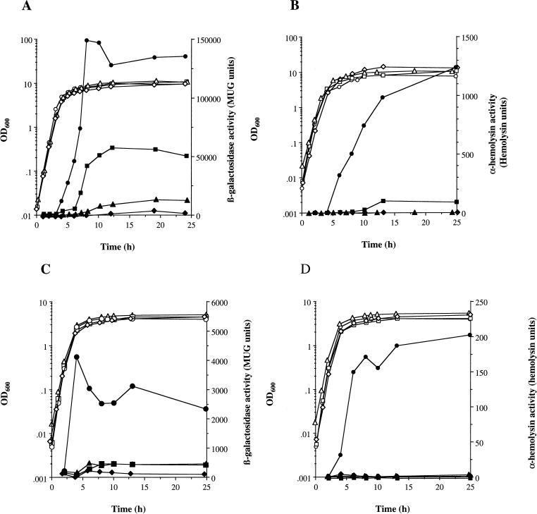
The staphylococcal accessory regulator (encoded by sarA) is an important global regulator of virulence factor biosynthesis in Staphylococcus aureus. To further characterize its role in virulence determinant production, an sarA knockout mutant was created by insertion of a kanamycin antibiotic resistance cassette into the sarA gene. N-terminal sequencing of exoproteins down-regulated by sarA identified several putative proteases, including a V8 serine protease and a novel metalloprotease, as the major extracellular proteins repressed by sarA. In kinetic studies, the sarA mutation delays the onset of alpha-hemolysin (encoded by hla) expression and reduces levels of hla to approximately 40% of the parent strain level. Furthermore, SarA plays a role in signal transduction in response to microaerobic growth since levels of hla were much lower in a microaerobic environment than after aerobic growth in the sarA mutant. An exoprotein exhibiting hemolysin activity on sheep blood, and up-regulated by sarA independently of the accessory gene regulator (encoded by agr), was specifically induced microaerobically. Transcriptional gene fusion and Western analysis revealed that sarA up-regulates both toxic shock syndrome toxin 1 gene (tst) expression and staphylococcal enterotoxin B production, respectively. This study demonstrates the role of sarA as a signal transduction regulatory component in response to aeration stimuli and suggests that sarA functions as a major repressor of protease activity. The possible role of proteases as regulators of virulence determinant stability is discussed.


Figures

FIG. 1
FIG. 1
Effects of regulatory mutations on exoprotein production in S. aureus SDS–10% (wt/vol) polyacrylamide gels show total extracellular protein production during growth as described in Materials and Methods. (A) S. aureus 8325-4 lanes 1 to 4 and PC1839 (sarA) (lanes 5 to 8) at T = 2, 4, 6, and 8 h, corresponding to log, early and late post-exponential, and stationary phases of growth, respectively. (B) S. aureus 8325-4 (lane 1), PC6911 (agr) (lane 2), PC1839 (sarA) (lane 3), and PC18391 (sarA agr) (lane 4), all in stationary phase (T = 8 h). All lanes contain exoproteins from the equivalent of 0.05 OD600 units of original culture. Representative sarA-regulated proteins are indicated by arrows. The molecular masses of protein standards (in kilodaltons) are shown.
FIG. 2
FIG. 2
Zymogram analysis of protease activity in exoproteins of S. aureus. Lane 1, S. aureus 8325-4; lane 2, PC6911 (agr); lane 3, PC1839 (sarA); lane 4, PC18391 (sarA agr). Exoproteins are from the equivalents of 0.05 (lanes 1 and 2) and 0.005 (lanes 3 and 4) OD600 units of original culture. Stationary-phase samples (T = 18 h) were prepared and separated on a 12% (wt/vol) polyacrylamide gel with casein as the substrate, and the gel was renatured as described in Materials and Methods. Proteolytic activity can be seen as a zone of clearing on the gel, indicated by arrows. The molecular masses of protein standards (in kilodaltons) are shown.
FIG. 3
FIG. 3
Western blot analysis of SEB production. (A) Western blot of total extracellular proteins of S. aureus S6 (lane 1), PC1700 (agr) (lane 2) PC1841 (sarA) (lane 3), and PC1845 (sarA agr) (lane 4). All lanes contain exoproteins from the equivalent of 0.05 OD600 units of original culture (T = 18 h). Proteins were separated by SDS-PAGE on a 10% (wt/vol) gel, transferred onto a nitrocellulose membrane (BDH), and probed with a 1:2,000 dilution of antibodies to SEB (Sigma) prior to colorimetric detection as described in Materials and Methods. (B) The relative intensity of each protein band quantified by densitometry (mean of three experiments plus standard deviation).
FIG. 4
FIG. 4
Role of sarA in the regulation of tst::lux expression during aerobic growth. S. aureus PC1072 (tst::lux) (○, •) and PC1091 (sarA tst::lux) (□, ■) were grown in BHI at 37°C with shaking at 250 rpm as described in Materials and Methods. Bacterial growth was measured by OD600 (○, □), and fusion expression was determined by luciferase activity (•, ■).
FIG. 5
FIG. 5
Role of sarA and agr in the regulation of hla::lacZ expression and Hla activity in response to environmental conditions. S. aureus PC322 (hla::lacZ) (○, •), PC3221 (sarA hla::lacZ) (□, ■), PC324 (agr hla::lacZ) (▵, ▴), and PC3241 (sarA agr hla::lacZ) (◊, ⧫) were grown in BHI at 37°C as described in Materials and Methods. (A and B) Aerobic growth in a shaking water bath at 250 rpm; (C and D) microaerobic growth on a shaking platform in a cabinet containing 8% O2, 5% CO2, and 87% N2. Bacterial growth was measured by OD600 (○, □, ▵, ◊). (A) hla::lacZ expression was determined by measuring β-galactosidase activity (•, ■, ▴, ⧫); (B) Hla activity in cell-free supernatants (•, ■, ▴, ⧫) was determined as described in Materials and Methods.
FIG. 6
FIG. 6
Nucleotide alignment of upstream regions of SarA-regulated genes of S. aureus. Alignments are based on homology of the noncoding regions 5′ of SarA-controlled genes to the proposed 29-bp SarA binding site (19) of the agr P2-P3 regulatory region (accession no. X52543 [50]). Best identity (58.6 to 72.4% identity over 29 bp to the agr sequence) was observed in AT-rich regions of tst (accession no. U93688), spa (accession no. J01786 [64]), hlb (accession no. X13404 [55]), seb (accession no. M11118 [37]), V8 protease (accession no. P04188 [11]), and hla (accession no. X01645 [28]). The distances of the hypothetical SarA binding site from transcriptional start (TS) sites are indicated where known. The 3′ ends of the tst, hlb, V8 protease, and hla sequences are located 145, 61, 9, and 270 bp upstream of their translational start sites. Nucleotides showing >50% identity over seven sequences are boxed in black. A predicted consensus sequence is indicated by upper- and lowercase letters representing 100 and >50% nucleotide identity, respectively, over the seven sequences.

References

    1. Abdelnour A, Arvidson S, Bremell T, Rydén C, Tarkowski A. The accessory gene regulator (agr) controls Staphylococcus aureus virulence in a murine arthritis model. Infect Immun. 1993;61:3879–3885. - PMC - PubMed
    1. Arvidson S. Studies on extracellular proteolytic enzymes from Staphylococcus aureus. II. Isolation and characterisation of an EDTA-sensitive protease. Biochim Biophys Acta. 1973;302:149–157. - PubMed
    1. Arvidson S O. Extracellular enzymes from Staphylococcus aureus. In: Easmon C S F, Adlam C, editors. Staphylococci and staphylococcal infections. Vol. 2. London, England: Academic Press Inc.; 1983. pp. 745–808.
    1. Ayora S, Götz F. Genetic and biochemical properties of an extracellular neutral metalloprotease from Staphylococcus hyicus subsp. hyicus. Mol Gen Genet. 1994;242:421–430. - PubMed
    1. Ayora S, Lindgren P-E, Götz F. Biochemical properties of a novel metalloprotease from Staphylococcus hyicus subsp. hyicus involved in extracellular lipase processing. J Bacteriol. 1994;176:3218–3223. - PMC - PubMed

Publication types

MeSH terms

LinkOut - more resources